نظرة عامة
البطين الأيمن ذو المخرجين مرض قلبي يكون موجودًا منذ الولادة. أي إنه عيب خلقي في القلب. في هذه الحالة المرضية، تحتوي حجرة القلب السفلية اليمنى على فتحتين لخروج الدم من القلب بدلاً من فتحة واحدة. ما يؤدي إلى تدفق جزء من الدم إلى الجسم بدلاً من الرئتين.
يكون لدى الرضّع الذين لديهم البطين الأيمن ذو المخرجين ثقب بين الحجرتين السفليتين للقلب. ويُطلق على هاتين الحجرتين البُطينَان. يُسمى هذا الثقب عيب الحاجز البُطيني. ويؤدي إلى اختلاط الدم المحمل بالأكسجين بالدم المفتقر إلى الأكسجين. ولذلك يمكن ألا يحصل الرضع الذين لديهم هذا الثقب على ما يكفي من الأكسجين في مجرى الدم. ويؤدي ذلك إلى تحول الجلد إلى اللون الرمادي أو الأزرق.
قد يكون البطين الأيمن ذو المخرجين مصحوبًا بأمراض قلبية أخرى موجودة منذ الولادة، مثل ثقوب القلب تغيّرات في صمامات القلب أو الأوعية الدموية.
البطين الأيمن مزدوج المخرج
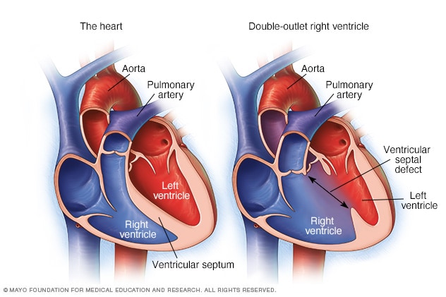
البطين الأيمن مزدوج المخرج
في حال البطين الأيمن ذي المخرجين، تحتوي الحجرة السفلية اليمنى (البطين الأيمن) على فتحتين لخروج الدم بدلاً من فتحة واحدة. وينتقل بعض الدم إلى الجسم بدلاً من الرئتين. ويكون هناك أيضًا ثقب بين حجرتي القلب السفليتين. ويُسمى هذا الثقب عيب الحاجز البُطيني. في القلب الطبيعي، كما هو موضح على اليسار، يتصل الشريان الرئوي بالبطين الأيمن، ويتصل الشريان الأورطي بالبطين الأيسر.
Types
There are several types of double-outlet right ventricle (DORV). The difference between these types is the location of the ventricular septal defect (VSD) and whether it is close to or far from the pulmonary artery and the aorta. The type of DORV determines which treatment may be right for your child.
Subaortic VSD
With subaortic VSD, the opening between the ventricles is close to the aorta. Because both arteries connect to the right ventricle in DORV, the left ventricle does not have its usual direct path to the aorta.
Subpulmonary VSD and Taussig-Bing type
With subpulmonary VSD, the opening between the ventricles is close to the pulmonary artery. Because of this, blood from the left ventricle tends to flow toward the pulmonary artery instead of the aorta. Taussig-Bing type is a form of DORV with this type of VSD.
Doubly committed VSD
With doubly committed VSD, the opening between the ventricles is close to both the aorta and the pulmonary artery. Because of its location, blood from the left ventricle can reach either artery.
Noncommitted VSD
With noncommitted VSD, also called remote VSD, the opening between the ventricles is farther from both the aorta and the pulmonary artery.
Fallot-type DORV
With Fallot-type (fa-LOW type) DORV, the VSD is usually close to the aorta or close to both the aorta and pulmonary artery. The pathway that carries blood from the right ventricle to the pulmonary artery may be narrowed. This narrowing lessens the amount of blood that can reach the lungs.
This is called Fallot-type DORV because it can resemble tetralogy of Fallot, which is another congenital heart defect that can lessen blood flow to the lungs.
المنتجات والخدمات
الأعراض
قد تشمل أعراض البُطين الأيمن ذي المخرجين ما يأتي:
- تصبغ الشفتين أو اللسان أو الأظافر بلون رمادي أو أزرق شاحب. ويختلف مدى سهولة ملاحظة تغيرات لون الجلد أو صعوبتها حسب لون الجلد.
- سرعة التنفس.
- مشاكل في التغذية والنمو وزيادة الوزن.
الحالات التي تستلزم زيارة الطبيب
إذا كنت تعتقد أن طفلك مصاب بأعراض أحد الأمراض القلبية، فحدد موعدًا طبيًا لإجراء فحص طبي على الفور.
الأسباب
السبب الدقيق للإصابة بحالة البُطين الأيمن ذي المخرجين غير معروف. تحدث هذه الحالة عندما يكون الجنين في طور النمو قبل الولادة.
هناك عوامل عدة قد تسهم في ظهورها، منها التغييرات الجينية أو بعض الأدوية أو الحالات المرَضية، أو العوامل البيئية أو تلك المرتبطة بنمط الحياة. كما أن التدخين أثناء الحمل قد يزيد من خطر إصابة الجنين ببعض عيوب القلب الخلقية.
عادة، يتصل الشريان الرئيسي في الجسم، المسمى الشريان الأورطي، بالحجرة السفلية اليسرى للقلب. بينما يتصل الشريان الرئوي بحجرة القلب السفلية اليمنى. لكن في حالات الأطفال الذين لديهم البطين الأيمن ذو المخرجين، يتصل الشريان الأورطي والشريان الرئوي جزئيًا أو كليًا بحجرة القلب السفلية اليمنى.
عوامل الخطورة
قد يؤدي وجود سيرة مَرضية عائلية للإصابة بأمراض القلب الخلقية إلى زيادة خطورة الإصابة بعيوب القلب الخلقية مثل البُطين الأيمن ذي المخرجين.
المضاعفات
تشمل مضاعفات البُطين الأيمن ذي المخرجين ضعف النمو وفشل القلب.