Overview

Double-outlet right ventricle (DORV) is a rare heart condition that is present at birth. A heart condition a child is born with is called a congenital heart defect. This is a change in the heart's structure that is present at birth.

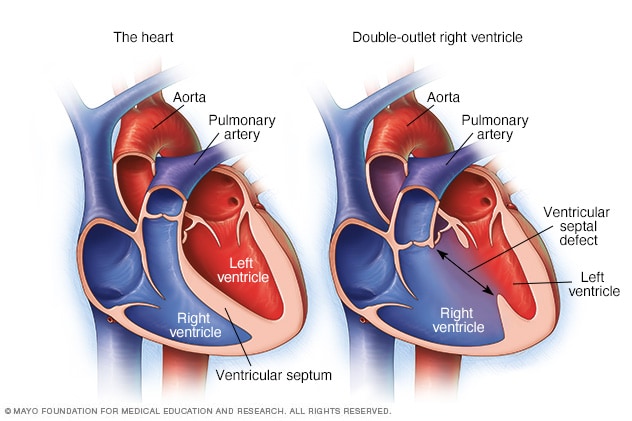
The heart has two lower chambers, called ventricles. Two large arteries carry blood away from the heart: the aorta and the pulmonary artery. The aorta connects to the left ventricle and carries blood to the body. The pulmonary artery connects to the right ventricle and carries blood to the lungs. A wall separates the two ventricles. In babies with double-outlet right ventricle, the aorta and the pulmonary artery connect to the right lower heart chamber, either partially or completely.

Most babies with DORV also have an opening in the wall between the two ventricles. This is called a ventricular septal defect (VSD).

DORV can look different from one child to another. Blood flow and the type of repair needed can be affected by:

  • The location of the VSD.
  • The position of the aorta and the pulmonary artery.
  • Narrowing in the pathways between the ventricles and the large arteries.

Most babies with DORV need surgery. The timing of surgery depends on your baby's heart structure, symptoms and blood flow. The healthcare team determines the type of treatment based on your child's heart structure.

With surgical treatment, most children have good long-term survival, although some may need additional heart procedures or lifelong follow-up.

Types

There are several types of double-outlet right ventricle (DORV). The difference between these types is the location of the ventricular septal defect (VSD) and whether it is close to or far from the pulmonary artery and the aorta. The type of DORV determines which treatment may be right for your child.

Subaortic VSD

With subaortic VSD, the opening between the ventricles is close to the aorta. Because both arteries connect to the right ventricle in DORV, the left ventricle does not have its usual direct path to the aorta.

Subpulmonary VSD and Taussig-Bing type

With subpulmonary VSD, the opening between the ventricles is close to the pulmonary artery. Because of this, blood from the left ventricle tends to flow toward the pulmonary artery instead of the aorta. Taussig-Bing type is a form of DORV with this type of VSD.

Doubly committed VSD

With doubly committed VSD, the opening between the ventricles is close to both the aorta and the pulmonary artery. Because of its location, blood from the left ventricle can reach either artery.

Noncommitted VSD

With noncommitted VSD, also called remote VSD, the opening between the ventricles is farther from both the aorta and the pulmonary artery.

Fallot-type DORV

With Fallot-type (fa-LOW type) DORV, the VSD is usually close to the aorta or close to both the aorta and pulmonary artery. The pathway that carries blood from the right ventricle to the pulmonary artery may be narrowed. This narrowing lessens the amount of blood that can reach the lungs.

This is called Fallot-type DORV because it can resemble tetralogy of Fallot, which is another congenital heart defect that can lessen blood flow to the lungs.

Symptoms

Signs and symptoms of DORV depend on how much blood flows from the heart to the lungs and to the rest of the body. Symptoms also depend on how much oxygen is in the blood.

Symptoms may start soon after birth or become more noticeable during the first weeks or months of life.

Symptoms may include:

  • Blue or gray skin, lips, or nail beds. This can happen when the blood has too little oxygen. The color change may be easier or harder to see depending on skin color.
  • Fast breathing or trouble breathing.
  • Not eating well.
  • Slow weight gain.
  • Low energy or unusual sleepiness.

When to see a doctor

If your baby has any of the symptoms of DORV, contact your healthcare professional right away.

Children with DORV also need regular checkups with a congenital heart doctor, called a cardiologist. This specialist treats heart conditions present at birth. These checkups are important even when your child feels well because DORV and the effects of earlier heart surgery can change over time.

When to seek emergency care

Seek emergency medical care if your baby:

  • Has severe trouble breathing.
  • Looks much more blue or gray than usual.
  • Is very sleepy or is hard to wake.

Causes

DORV develops before birth while the heart and large arteries are forming. The exact cause is not known. Changes in genes or chromosomes that happen before birth may affect how the heart develops and may play a role in DORV.

Risk factors

There is no single known risk factor for DORV. A family history of congenital heart disease or a known genetic condition may increase the chance of a baby having a congenital heart disease, such as DORV.

Other conditions and DORV

DORV can resemble another congenital heart condition called transposition of the great arteries. In this condition, the two main arteries leaving the heart connect to the opposite ventricles rather than to their usual positions. The aorta comes from the right ventricle, and the pulmonary artery comes from the left ventricle.

Babies with DORV also may have other health conditions, such as:

  • Heterotaxy. This is a congenital condition in which organs in the chest or abdomen are arranged differently from their usual positions.
  • Hypoplastic left heart syndrome. This is a congenital heart condition in which the left side of the heart is severely underdeveloped. Some babies with DORV also have a very small left ventricle and other heart differences that can block blood flow through the left side of the heart.

Complications

Complications can happen before surgery to repair DORV, such as:

  • Too little oxygen in the blood.
  • Too much blood flow to the lungs.
  • Damage to blood vessels in the lungs.
  • Slow growth.
  • Heart failure. Heart failure happens when the heart cannot pump enough blood to meet the body's needs.

Diagnosis

DORV may be found before birth during a routine ultrasound. Ultrasound uses sound waves to make images inside the body. If the ultrasound shows a possible heart issue, a fetal echocardiogram is done to confirm the diagnosis and provide more details. The results also can help the healthcare team plan care around delivery.

Fetal echocardiogram

A fetal echocardiogram is a detailed ultrasound of a baby's heart before birth. It can show the heart chambers, the VSD, the aorta and pulmonary artery. It also can show whether there is narrowing in the pathways that carry blood from the heart or other differences in the heart's structure.

Echocardiogram

After birth, an echocardiogram is the main imaging test used to diagnose DORV and look closely at the heart's structure. An echocardiogram uses sound waves to make moving pictures of the heart. It can show where the VSD is and how the aorta and pulmonary artery connect to the heart. It also can show how well the ventricles pump blood and how well the heart valves work. Heart valves are like doors that open only one way to let blood flow forward and close to keep it from flowing backward. An echocardiogram also can show narrowing in the pathways that carry blood from the heart.

CT scan

A cardiac CT scan uses X-rays to create detailed pictures of the heart. This test can provide 3D images when an echocardiogram does not show enough detail about the heart's structure.

MRI

MRI uses a magnetic field and radio waves to create detailed pictures of the heart. MRI can provide more information about the heart's structure and how well it works. It may be used to look at the heart after treatment.

Cardiac catheterization

Cardiac catheterization is a procedure in which a thin, flexible tube called a catheter is guided through a blood vessel to the heart. Not every child with DORV needs this procedure for diagnosis. The care team may use it to measure pressure and oxygen levels in the heart, see how blood flows through the heart, or treat certain heart problems.

Treatment

Treatment for DORV aims to improve blood flow from the heart to the body and lungs and open narrowed pathways. When possible, treatment also creates separate pathways so oxygen-rich blood flows to the body. Oxygen-poor blood flows to the lungs to help it pick up more oxygen.

Some newborns need treatment soon after birth to improve blood flow while the care team plans surgery. Prostaglandin E1 is a medicine that is given through an IV. It keeps a blood vessel called the ductus arteriosus open. This blood vessel is open before birth and usually closes soon after birth. In some babies with DORV, keeping it open allows more blood to reach the lungs or the rest of the body while the care team makes a treatment plan.

Depending on the heart structure, a baby also may need a procedure called atrial septostomy. This procedure uses a thin, flexible tube called a catheter to make or enlarge an opening between the two upper chambers of the heart. The opening allows blood to mix between these chambers, which can help increase the amount of oxygen in the blood that reaches the body. Some babies may need other procedures or an earlier surgery before DORV can be repaired.

Surgery

Most babies with DORV need surgery to improve how blood flows from the heart to the lungs and the rest of the body. The type of surgery your child needs depends on your child's heart structure.

The surgical team looks at:

  • Where the VSD is located.
  • The positions of the aorta and the pulmonary artery.
  • The size of both ventricles and how well they pump.
  • How well the heart valves work.
  • Narrowing in the pathways that carry blood from the heart.
  • Blood vessels that supply the heart muscle, called the coronary arteries.

These details help the team decide what type of repair is possible and right for your child.

Intraventricular tunnel repair

This surgery may be done when the VSD is close to the aorta. During an intraventricular tunnel repair, a pathway is created inside the heart from the left ventricle through the VSD and into the aorta. This allows the left ventricle to pump blood through the aorta to the body.

If the pathway from the right ventricle to the pulmonary artery is narrow, it also may need to be widened or rebuilt.

Arterial switch operation

An arterial switch operation may be part of the repair procedure for babies with Taussig-Bing type or other types of DORV in which the VSD is close to the pulmonary artery. During this surgery, a surgeon moves the aorta and pulmonary artery and creates a pathway through the VSD to direct blood from the left ventricle to the aorta. This allows the left ventricle to pump blood through the aorta to the body. It also allows the right ventricle to pump blood through the pulmonary artery to the lungs.

The blood vessels that supply the heart muscle, called the coronary arteries, also are moved to the aorta.

Rastelli procedure

The Rastelli procedure may be used when a pathway can be made from the left ventricle to the aorta, but the pathway from the right ventricle to the pulmonary artery is too narrow. This may happen when the valve that controls blood flow from the right ventricle to the pulmonary artery is narrowed. This is called pulmonary valve stenosis.

During the Rastelli procedure, a surgeon creates a pathway from the left ventricle through the VSD and into the aorta. This allows the left ventricle to pump blood to the body. A tube with a valve inside, called a conduit, is placed between the right ventricle and the pulmonary artery. The conduit is the pathway that allows blood to flow from the right ventricle to the pulmonary artery so the right ventricle can pump blood to the lungs.

The conduit does not grow as a child grows. Over time, it can narrow, harden or leak. Another catheter procedure or surgery may be needed to repair or replace the conduit.

Single-ventricle surgery options

Sometimes, the heart's structure does not allow both ventricles to work separately. When this happens, the care team may consider what is called a single-ventricle pathway. This is a more complex treatment that involves several surgeries done in stages to change how blood moves through the heart and lungs. After these surgeries, one ventricle does most of the heart's pumping work.

A single-ventricle pathway may be considered when:

  • One ventricle is very small.
  • A heart valve has complex attachments.
  • The VSD is too far from the aorta or pulmonary artery to create the pathway needed for a two-ventricle repair.

Other complex types of repairs

Other, more complex types of surgery may be done in rare situations based on the unique features of DORV in some babies. Cardiologists and surgeons recommend the surgery that is right for each child.

Sept. 10, 2026

Living with double-outlet right ventricle?

Connect with others like you for support and answers to your questions in the Heart & Blood Health support group on Mayo Clinic Connect, a patient community.

Heart & Blood Health Discussions

teatime
Stopping Carvedilol (Coreg): When will the effects wear off?

415 Replies Tue, Sep 08, 2026

pop77
Living with high calcium score

118 Replies Sat, Sep 12, 2026

sjy70
High calcium score: I'm in shock

241 Replies Wed, Aug 26, 2026

See more discussions
  1. Xu Z, et al. Double outlet ventricles: Review of anatomic and imaging characteristics. Heart. 2023; doi:10.1136/heartjnl-2022-321955.
  2. Bell-Cheddar Y, et al. Double outlet right ventricle. Frontiers in Pediatrics. 2023; doi:10.3389/fped.2023.1244558.
  3. Holten-Andersen M, et al. Current outcomes of live-born children with double outlet right ventricle in Norway. European Journal of Cardio-Thoracic Surgery. 2023; doi:10.1093/ejcts/ezac560.
  4. Gurvitz M, et al. 2025 ACC/AHA/HRS/ISACHD/SCAI guideline for the management of adults with congenital heart disease: A report of the American College of Cardiology/American Heart Association Joint Committee on Clinical Practice Guidelines. Circulation. 2025; doi:10.1161/CIR.0000000000001402.
  5. Pang K, et al. Long-term surgical outcomes in double outlet right ventricle based on detailed anatomical sub-typology. European Journal of Cardio-Thoracic Surgery. 2025; doi:10.1093/ejcts/ezaf334.
  6. Rickert-Sperling S, et al., eds. Congenital Heart Diseases: The Broken Heart: Clinical Features, Human Genetics and Molecular Pathways. 2nd ed. Springer; 2024. https://link.springer.com/book/10.1007/978-3-031-44087-8. Accessed Aug. 17, 2026.
  7. Öcal A, et al. Navigating the spectrum of double-outlet right ventricle presentations: Outcomes from a contemporary cohort based on subtypes. Echocardiography. 2024; doi:10.1111/echo.15790.
  8. Moon-Grady AJ, et al. Guidelines and recommendations for performance of the fetal echocardiogram: An update from the American Society of Echocardiography. Journal of the American Society of Echocardiography. 2023; doi:10.1016/j.echo.2023.04.014.
  9. Pontailler M, et al. Long-term results after the réparation à l'étage ventriculaire procedure for transposition of the great arteries and double-outlet right ventricle with pulmonary stenosis. European Journal of Cardio-Thoracic Surgery. 2023; doi:10.1093/ejcts/ezad409.
  10. Lacour-Gayet F, et al. Multicentre study on late outcomes of biventricular repair of double outlet right ventricle. European Journal of Cardio-Thoracic Surgery. 2024; doi:10.1093/ejcts/ezad423.
  11. Medical review (expert opinion). Mayo Clinic. Sept. 3, 2026.

Related

Associated Procedures

Products & Services