Overview
Echocardiogram
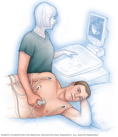
Echocardiogram
An echocardiogram uses sound waves to show how blood flows through the heart and heart valves. Sensors attached to the chest and sometimes the legs check the heart rhythm during the test. The test can help a healthcare professional diagnose heart conditions.
An echocardiogram uses sound waves to create pictures of the heart. This common test can show blood flow through the heart and heart valves. Your healthcare professional can use the pictures from the test to find heart disease and other heart conditions.
An echocardiogram also is called an echo or echocardiography. You may hear several other terms used for the same type of heart ultrasound:
- Cardiac echo.
- Cardiac ultrasound.
- Echo cardiogram.
- Echo ultrasound.
There are different types of echocardiograms. The type you have depends on the reason for the test and your overall health. Some types of echocardiograms are done during exercise or pregnancy.
An echocardiogram is different from an electrocardiogram (ECG or EKG). An echocardiogram uses ultrasound to create pictures of the heart and blood flow. An ECG or EKG records the heart's electrical activity.
Products & Services
Why it's done
Your healthcare professional may use an echocardiogram to look at your heart. The test shows how blood moves through the heart chambers and heart valves. Your healthcare professional may recommend this test if you have chest pain or shortness of breath.
An echocardiogram can help your healthcare professional check for:
- Heart failure. The test can show how well the heart pumps and fills with blood.
- Heart muscle damage. The test can show changes in how the heart wall moves that may be related to a heart attack or reduced blood flow.
- The cause of a heart murmur. This includes changes to heart valves.
- Heart valve disease. This may include a valve that leaks, called regurgitation, or a valve that has narrowed, called stenosis.
- Heart conditions present at birth. These are called congenital heart defects and include openings between heart chambers.
A standard echocardiogram doesn't directly show if you have a blockage of a heart artery. A stress echocardiogram can help identify partial or complete blockages by showing how the heart muscle moves when the heart is working harder. To confirm a blockage, you may need more imaging of the heart arteries, either with a coronary angiogram or a CT coronary angiogram.
Types of echocardiogram
The type of echocardiogram you have depends on the information your healthcare professional needs.
Transthoracic echocardiogram
A transthoracic echocardiogram (TTE) is a standard echocardiogram. It also is called a heart ultrasound. It's noninvasive, so the wand used to make pictures stays outside of the chest.
The test shows how blood flows through the heart and heart valves. You may receive contrast through an IV. The contrast helps the heart's structures show up better on the images.
Transesophageal echocardiogram
Your healthcare professional may recommend a transesophageal echocardiogram (TEE) if a standard echocardiogram doesn't provide enough detail. A TEE creates detailed pictures of the heart and the body's main artery, called the aorta. A TEE creates these pictures using a probe that goes through your mouth into your esophagus, which is the tube that connects the throat to the stomach.
This test may not be safe if you have active bleeding in the upper digestive tract or a tumor or tear in the esophagus.
Fetal echocardiogram
During pregnancy, a fetal echocardiogram checks the unborn baby's heart. It's noninvasive. A healthcare professional moves an ultrasound wand over your abdomen to create the pictures. The test doesn't use X-rays.
Stress echocardiogram
A stress echocardiogram is done with intense exercise to check how the heart responds to physical activity. A healthcare professional takes baseline images before you start to exercise, then more after you exercise.
Your healthcare professional may order this test if they think the arteries that supply your heart may be narrowed or blocked. If you can't exercise, you may be given medicine that affects the heart like exercise does.
Echocardiogram methods
Your healthcare team may use several imaging methods during an echocardiogram:
Two-dimensional (2D) and three-dimensional (3D) echocardiogram
These methods show the heart walls, valves and large blood vessels connected to your heart. A 2D echocardiogram is the standard test. Some medical centers and hospitals offer 3D echocardiograms. The images can provide more details about the lower left heart chamber, the heart's main pumping area.
Doppler echocardiogram
Sound waves change pitch when they bounce off blood cells moving through the heart and blood vessels. These changes are called Doppler signals. A doppler echocardiogram measures the speed and direction of blood flow within the heart and vessels. It can help show blocked or leaking valves and estimate pressure in the artery that carries blood from your heart to your lungs, called the pulmonary artery.
Color flow imaging
This shows the blood flow in the heart in color. It helps find leaky heart valves and other changes in blood flow. Most of the time, red shows blood moving toward the ultrasound wand and blue shows blood moving away from it.
Bubble study
During some echocardiograms, a healthcare professional injects a saltwater solution, called saline, that contains tiny bubbles. If the bubbles cross from the right side of the heart to the left, that means blood is moving through an opening between the two sides of the heart. The movement could mean there's a hole in the wall between heart chambers called a septal defect.
More Information
Risks
Echocardiography uses sound waves, called ultrasound. Medical experts generally consider ultrasound safe. There is no X-ray exposure.
Other risks depend on the type of echocardiogram you have.
A standard transthoracic echocardiogram usually doesn't hurt, though you may feel some discomfort when the ultrasound wand pushes against your chest. The healthcare professional may need to press firmly to create the clearest pictures of your heart.
There may be a small risk of a reaction to the contrast used for some echocardiograms. Some people get backaches, headaches or rashes. Severe reactions are very rare.
If you have a transesophageal echocardiogram, your throat may be sore afterward. Rarely, the probe used for this test can injure the throat or esophagus. Other risks of a TEE include:
- Difficulty swallowing.
- Weak or scratchy voice.
- Muscle spasms in the throat or lungs.
- Minor bleeding in the throat or esophagus.
- Injury to teeth, gums or lips.
- A hole in the esophagus, called esophageal perforation.
- A heartbeat that is not regular, called an arrhythmia.
- Nausea from medicines used during the test.
Some stress echocardiograms use medicine instead of exercise. The medicine may temporarily cause a fast or irregular heartbeat, a flushed feeling or low blood pressure. An allergic reaction also is possible. Serious complications, such as heart attacks, are rare.
How you prepare
How you prepare for an echocardiogram depends on the type you're having. If you're having a transesophageal echocardiogram, you usually receive medicine to help you relax. Arrange for a ride home, and plan to miss work for the rest of the day.
Food and medicines
Before your test appointment, ask your healthcare professional if you can take your medicines as usual. Make sure your healthcare team knows about all the medicines you take, including those bought without a prescription.
You can often eat or drink as usual before a standard transthoracic echocardiogram. Most types of echo do not need special preparation.
If you're having a transesophageal echocardiogram, which takes pictures from inside your esophagus, you may be told not to eat or drink for several hours before the test. Your healthcare professional gives you details on how to prepare.
Caffeine can affect some medicines used for certain stress tests. If you're having a stress echocardiogram that uses medicine, your care team may recommend avoiding caffeine for 12 to 24 hours before the test. This may include avoiding coffee, tea, cocoa, chocolate, some energy drinks and caffeinated sodas. Follow your care team's preparation instructions.
What you can expect
Before the procedure
You may have an echocardiogram at a medical center, hospital or a healthcare professional's office.
The procedure is generally the same for men and women. For most types of echocardiograms, your care team asks you to remove clothing from your upper body and change into a gown.
An electrocardiogram (ECG or EKG) is often done at the same time as an echocardiogram. A healthcare professional attaches sticky patches to your chest. The healthcare professional may put patches on your legs too. The patches contain sensors, called electrodes, that track your heartbeat.
What happens next depends on the type of echocardiogram you're having.
During the procedure
During an echocardiogram, your care team monitors your health. You can tell them at any time if you feel discomfort or have concerns.
Transthoracic echocardiogram
This is the standard type of echocardiogram. It takes pictures of the heart from outside the body.
During a standard echocardiogram:
- A healthcare professional puts gel on the ultrasound wand, called a transducer. The gel helps transmit the sound waves.
- The healthcare professional presses the wand against the skin and moves it over the chest area.
- The wand sends sound waves through the chest to the heart.
- The wand records the sound waves that bounce back from the heart.
- A computer turns the information into moving images that appear right away on a monitor.
During the test, your healthcare professional may ask you to breathe in a certain way or to roll onto your left side. If the images are not clear enough, your team may give you contrast by IV to help the heart's structures show up more clearly. You also may receive by IV saline with tiny gas bubbles to check for an opening between heart chambers.
A fetal echocardiogram is similar to the standard test, except the healthcare professional moves the wand over the abdomen.
Transesophageal echocardiogram
This type of echocardiogram provides more-detailed images of the heart and heart valves. Unlike a standard echocardiogram, this test takes pictures from inside the body.
This test uses medicines called sedatives to help you relax. Before the test starts, your healthcare team:
- Inserts an IV into your arm or hand to deliver medicine and contrast.
- Numbs your throat with a spray or gel to help control your gag reflex.
- Applies small patches called electrodes to your chest and possibly legs to track your heartbeat.
- Places a face mask or a small tube placed in your nostrils to provide extra oxygen.
During a transesophageal echocardiogram:
- The healthcare professional guides a thin, flexible ultrasound probe into your mouth. The probe moves down your throat into your esophagus, near your heart. The healthcare professional may ask you to swallow as the probe moves down.
- A transducer at the end of the probe sends sound waves to your heart and records the echoes that bounce back. A computer turns the echoes into moving images on a monitor.
- Your team watches your oxygen level and blood pressure during the test.
Stress echocardiogram
A stress echocardiogram is similar to a standard transthoracic echocardiogram, but your healthcare professional takes images before and after exercise. Some heart conditions cause changes only during physical activity.
A stress echocardiogram often involves walking on a treadmill or riding a stationary bike while an ECG records your heart's electrical activity. The healthcare professional takes images of your heart before and after you exercise. If you can't exercise, your healthcare professional may give you medicine that affects the heart like exercise does.
After the procedure
Most echocardiograms take less than an hour. If you have a transesophageal echocardiogram, your care team may watch you until you are fully awake and ready to go home.
After the test, a healthcare professional reviews the images. You may be asked to wait in case more images are needed.
After a standard echocardiogram, you can usually return to your daily activities. If you had a transesophageal echocardiogram with sedation, follow the instructions you receive about eating, drinking and driving.
If the results don't show anything that needs a closer look, you may not need more tests. If they do, you may need more testing. Your healthcare professional may refer you to a heart doctor, called a cardiologist, if you aren't already seeing one.
Results
After the procedure, a doctor trained in echocardiography reviews the images to determine what they show. Results are often ready by the next business day.
Information from an echocardiogram may show:
- Changes in heart size. Weakened or damaged heart valves, high blood pressure, or other diseases can cause thickened heart walls or enlarged heart chambers.
- Pumping strength. An echocardiogram can show the heart's ejection fraction (EF). EF is the percentage of blood the left ventricle pumps out with each contraction. The calculation compares the amount of blood in the chamber before it pumps with the amount left afterward. The test also shows how much blood the heart pumps in one minute. This is called cardiac output.
- Heart muscle damage. The test can show how the heart wall moves. Areas of the heart wall that move weakly may be damaged. Such damage might be due to a lack of oxygen or a heart attack.
- Heart valve disease. An echocardiogram can show how the heart valves open and close. The test is often used to check for leaky heart valves. It can help diagnose valve disease, such as heart valve regurgitation and valve stenosis.
- Heart conditions present at birth, called congenital heart defects. An echocardiogram can show changes in the structure of the heart and heart valves. The test also is used to look for changes in the connections between the heart and major blood vessels.
Clinical trials
Explore Mayo Clinic studies of tests and procedures to help prevent, detect, treat or manage conditions.
Aug. 21, 2026