Tetralogy of Fallot Symptoms and causes
Overview
Tetralogy of Fallot (teh-TRAL-uh-jee of fuh-LOW) is a rare heart condition that is present at birth. That means it's a congenital heart defect. A baby born with Tetralogy of Fallot has four different heart conditions.
The four heart conditions affect the structure of the heart. As a result, there are changes in blood flow through the heart and to the rest of the body. Babies with tetralogy of Fallot often have blue or gray skin color due to low oxygen levels.
Tetralogy of Fallot is usually diagnosed during pregnancy or soon after a baby is born. If the heart changes and symptoms are mild, tetralogy of Fallot may not be noticed or diagnosed until adulthood.
Babies with tetralogy of Fallot need surgery to fix the heart. They also need regular health checkups for life.
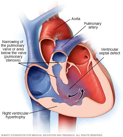
Tetralogy of Fallot
Tetralogy of Fallot is a combination of four heart changes present at birth. There is a hole in the heart called a ventricular septal defect. There also is a narrowing of the pulmonary valve or other area along the pathway between the heart and lungs. Narrowing of the pulmonary valve is called pulmonary stenosis. The body's main artery, called the aorta, is misplaced. The lower right heart chamber wall is thickened, a condition called right ventricular hypertrophy. Tetralogy of Fallot changes how blood flows through the heart and to the rest of the body.
Symptoms
Tetralogy of Fallot symptoms depend on how much blood flow is blocked from leaving the heart to go to the lungs. Symptoms may include:
- Blue or gray skin color. Depending on skin color, these changes may be harder or easier to see.
- Shortness of breath and rapid breathing, especially during feeding or exercise.
- Trouble gaining weight.
- Getting tired easily during play or exercise.
- Crankiness.
- Crying for long periods of time.
- Fainting.
Tet spells
Some babies with tetralogy of Fallot suddenly develop deep blue or gray skin, nails and lips. This usually happens when the baby cries, eats or is upset. These episodes are called tet spells.
Tet spells are caused by a rapid drop in the amount of oxygen in the blood. They are most common in young infants, around 2 to 4 months old. Tet spells may not be as easy to see in toddlers and older children. That's because toddlers and older children typically squat when they're short of breath. Squatting sends more blood to the lungs.
When to see a doctor
Serious congenital heart conditions such as tetralogy of Fallot are often diagnosed before or soon after a baby is born. Seek medical help if you notice that your baby has these symptoms:
- Trouble breathing.
- Blue or gray color of the skin.
- Lack of alertness.
- Seizures.
- Weakness.
- More cranky than usual.
If your baby's skin becomes blue or gray, place your baby on the side and pull the baby's knees up to the chest. This helps increase blood flow to the lungs. Call 911 or your local emergency number right away.
Causes
Tetralogy of Fallot happens during pregnancy when the baby's heart grows. Usually, the cause isn't known.
Tetralogy of Fallot includes four changes in heart structure:
- Narrowing of the valve between the heart and the lungs, called pulmonary valve stenosis. This condition reduces blood flow from the heart to the lungs. The narrowing may just involve the valve. Or it could happen in more than one place along the pathway between the heart and lungs. Sometimes the valve isn't formed. Instead, a solid sheet of tissue blocks blood flow from the right side of the heart. This is called pulmonary atresia.
- A hole between the bottom heart chambers, called a ventricular septal defect. A ventricular septal defect changes how blood flows through the heart and lungs. Oxygen-poor blood in the lower right chamber mixes with oxygen-rich blood in the lower left chamber. The heart has to work harder to pump blood through the body. The problem may weaken the heart over time.
- Shifting of the body's main artery. The body's main artery is called the aorta. It's usually attached to the left lower heart chamber. In tetralogy of Fallot, the aorta is in the wrong place. It's shifted to the right and sits directly above the hole in the heart wall. This changes how blood flows from the aorta to the lungs.
- Thickening of the right lower chamber of the heart, called right ventricular hypertrophy. When the heart has to work too hard, the wall of the right lower heart chamber gets thick. Over time, this may cause the heart to become weak and stop pumping blood as well as it should.
Some people with tetralogy of Fallot have other changes that affect the aorta or heart arteries. There also may be a hole between the heart's upper chambers, called atrial septal defect.
Risk factors
The exact cause of tetralogy of Fallot isn't known. Some things may raise the risk of a baby being born with tetralogy of Fallot. Risk factors include:
- Family history of the condition.
- Having a virus during pregnancy. This includes rubella, also known as German measles.
- Drinking alcohol during pregnancy.
- Eating poorly during pregnancy.
- Smoking during pregnancy.
- Being older than 35 during pregnancy.
- Down syndrome or DiGeorge syndrome in the baby.
Complications
Without treatment, tetralogy of Fallot usually leads to life-threatening complications. The complications may cause disability or death by early adulthood.
A possible complication of tetralogy of Fallot is infection of the inner lining of the heart or heart valves. This is called infective endocarditis. Sometimes medicines called antibiotics are given before dental work to prevent this type of infection. Ask your healthcare team if preventive antibiotics are right for you or your baby.
Complications also are possible after surgery to repair tetralogy of Fallot. But most people do well after such surgery. When complications occur, they may include:
- Backward flow of blood through a heart valve.
- Irregular heartbeats.
- A hole in the heart that doesn't go away after surgery.
- Changes in the size of the heart chambers.
- Swelling of part of the aorta, called aortic root dilation.
- Sudden cardiac death.
Another treatment or surgery may be needed to fix these complications.
Congenital heart defects and pregnancy
People born with a complex congenital heart defect may be at risk of complications during pregnancy. Talk with your healthcare team about the possible risks and complications of pregnancy. Together you can discuss and plan for any special care needed.
Prevention
Because the exact cause of most congenital heart defects isn't known, it may not be possible to prevent these conditions. If you have a high risk of giving birth to a child with a congenital heart defect, genetic testing and screening may be done during pregnancy.
There are some steps you can take to help lower your child's overall risk of birth defects, such as:
- Get proper prenatal care. Regular checkups with a healthcare team during pregnancy can help keep the pregnant parent and baby healthy.
- Take a multivitamin with folic acid. Taking 400 micrograms of folic acid daily can lower the risk of birth defects in the brain and spinal cord. It may help lower the risk of heart defects as well.
- Don't drink or smoke. These lifestyle habits can harm a baby's health. Also stay away from secondhand smoke.
- Get a rubella vaccine, also called a German measles vaccine. An infection with the rubella virus during pregnancy may affect a baby's heart development. Get vaccinated before trying to get pregnant.
- Manage blood sugar. If you have diabetes, managing your blood sugar can lower the risk of congenital heart defects.
- Manage long-term health conditions. If you have other health conditions, including a rare inherited condition called phenylketonuria, talk with your healthcare team. Ask about the best ways to treat and manage them.
- Stay away from harmful substances. During pregnancy, have someone else do any painting and cleaning with strong-smelling products.
- Check with your healthcare team before taking any medicines. Some medicines can cause birth defects. Tell your healthcare team about all the medicines you take, including those bought without a prescription.

Top ranked in the U.S.
Mayo Clinic in Rochester, Minnesota, has been recognized as one of the top Cardiology & Heart Surgery hospitals in the nation by U.S. News & World Report.
CON-20316969
Mayo Clinic does not endorse companies or products. Advertising revenue supports our not-for-profit mission.
Advertising & Sponsorship
Mayo Clinic Press
Check out these best-sellers and special offers on books and newsletters from Mayo Clinic Press.
