Diagnosis
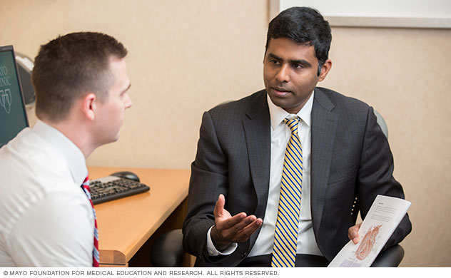 Ventricular tachycardia consultation at Mayo Clinic
A thorough physical exam, medical history and testing are required to diagnose ventricular tachycardia.
Ventricular tachycardia sometimes requires emergency medical care and may be diagnosed at a hospital. When possible, a healthcare professional may ask you or your family questions about symptoms, lifestyle habits and medical history.
Tests
Electrocardiogram
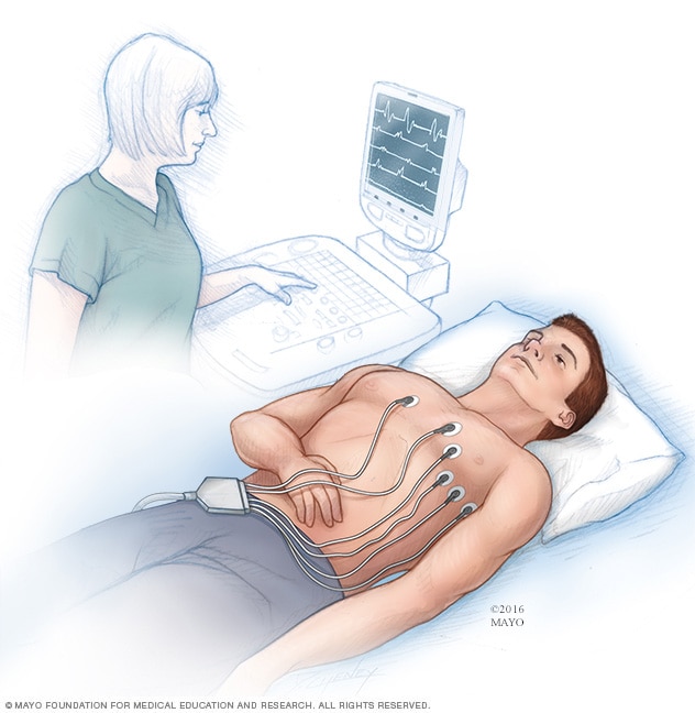
Electrocardiogram
An electrocardiogram (ECG or EKG) is a test to record the electrical signals in the heart. It shows how the heart is beating. Sticky patches called electrodes are placed on the chest and sometimes on the arms or legs. Wires connect the patches to a computer, which prints or displays results.
Holter monitor
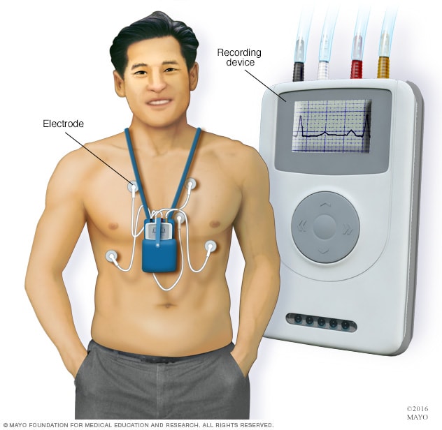
Holter monitor
A Holter monitor is a small, wearable device that continuously records the heart's rhythm for a day or more. A healthcare professional can review the data captured on the recording device to determine if an irregular heartbeat, called an arrhythmia, is found.
Cardiac event monitor
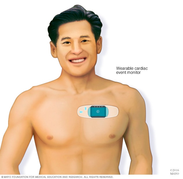
Cardiac event monitor
A wearable cardiac event monitor may be used to diagnose tachycardia. This type of portable ECG device records heart activity only during episodes of irregular heartbeats, called arrhythmias.
Tests are done to check the heart and confirm a diagnosis of ventricular tachycardia, also called V-tach or VT. Test results also can help determine if another health problem is causing V-tach.
- Electrocardiogram (ECG or EKG). This is the most common test to diagnose tachycardia. An ECG shows how the heart is beating. Small sensors, called electrodes, attach to the chest and sometimes the arms and legs. Wires connect the sensors to a computer, which prints or displays results. The test can help determine the type of tachycardia.
- Holter monitor. If a standard ECG doesn't give enough details, your care team may ask you to wear a heart monitor at home. A Holter monitor is a small ECG device. It is worn for a day or more to record the heart's activity during daily activities. Some personal devices, such as smartwatches, offer portable ECG monitoring. Ask your care team if this is an option for you.
- Event monitor. This portable ECG device is worn for up to 30 days or until you have an irregular heartbeat or symptoms. You typically press a button when symptoms occur.
- Implantable loop recorder. This small device records the heartbeat continuously for up to three years. It's also called a cardiac event recorder. The device tells your care team how your heart beats during daily activities. It is placed just under the skin of the chest during a minor procedure.
Exercise stress test
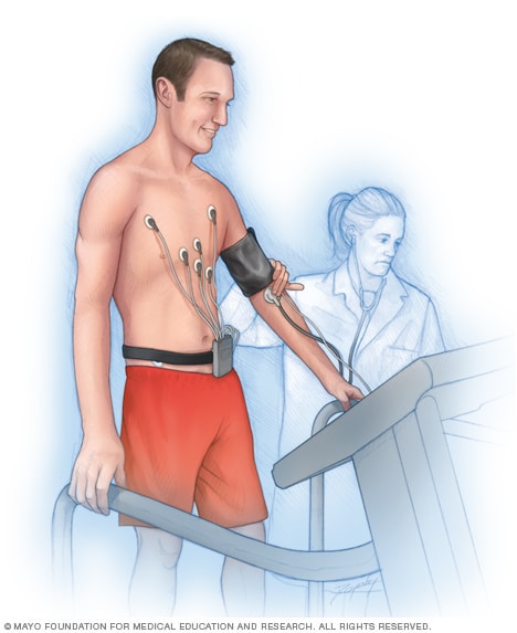
Exercise stress test
In an exercise stress test, sensors called electrodes are placed on the chest and sometimes the arms and legs. The sensors record information about the heartbeat. A healthcare professional checks the heart while the person walks on a treadmill or pedals a stationary bike.
Imaging tests can help your care team check the structure of your heart. Cardiac imaging tests used to diagnose ventricular tachycardia include:
- Chest X-ray. A chest X-ray shows the condition of the heart and lungs.
- Echocardiogram. This test is an ultrasound of the heart. It uses sounds waves to create a picture of the beating heart. It can show areas of poor blood flow and heart valve problems.
- Exercise stress test. This isn't an imaging test, but it may be done during an imaging test called an echocardiogram. The test usually involves walking on a treadmill or riding a stationary bike while a care professional watches the heartbeat. Some types of tachycardia are triggered or worsened by exercise. If you can't exercise, you may get medicine that affects the heartbeat like exercise does.
- Cardiac magnetic resonance imaging (MRI). This test creates still or moving pictures of blood flow through the heart. It is most often done to determine a cause of ventricular tachycardia or ventricular fibrillation.
- Cardiac computerized tomography (CT). CT scans combine several X-ray images to provide a more detailed view of the area being studied. A CT scan of the heart, called a cardiac CT scan, may be done to find the cause of ventricular tachycardia.
- Coronary angiogram. A coronary angiogram is done to check for blocked or narrowed blood vessels in the heart. It uses a dye and special X-rays to show the inside of the coronary arteries. This test may be done to look at the heart's blood supply in people who have ventricular tachycardia or ventricular fibrillation.
Cardiac MRI animation
Watch how heart MRI, also called cardiac MRI, is used to view the heart.
Other tests are done to confirm tachycardia and its cause and to learn how it leads to other health concerns. These tests include:
- Electrophysiological (EP) study. An EP study is a series of tests that help create a very detailed map of how signals move between each heartbeat. It may be done to confirm tachycardia or to find where in the heart the faulty signaling occurs. It's usually done to diagnose isolated irregular heartbeats. A doctor inserts one or more thin, flexible tubes into a blood vessel and guides them to the heart. Sensors on the tips of the tubes send electrical signals to the heart and record the heart's electrical activity.
- Tilt table test. A tilt table test may be done to better understand how tachycardia leads to fainting. A healthcare professional checks your heart rate and blood pressure as you lie flat on a table. Straps secure you to the table. Then, under careful supervision, the table slowly tilts until you are standing. The test shows how the heart and nervous system respond to changes in body position.
More Information
Treatment
 Ventricular tachycardia treatment at Mayo Clinic
A Mayo Clinic healthcare professional delivers specialized care for ventricular tachycardia.
Ventricular tachycardia that lasts longer than 30 seconds, called sustained V-tach, needs emergency medical treatment. Sustained V-tach may sometimes lead to sudden cardiac death.
The goals of ventricular tachycardia treatment are to:
- Slow a rapid heartbeat.
- Prevent future episodes of a fast heartbeat.
Ventricular tachycardia treatment may include medicines, procedures and devices to control or reset the heart rhythm, and heart surgery.
If another medical condition is causing tachycardia, treating the underlying problem may reduce or prevent episodes of a fast heartbeat.
Medications
Medicines are given to slow the fast heart rate. Medicines used to treat tachycardia may include beta blockers. You may need more than one medicine. Talk to your healthcare team about the type of medicine that is best for you.
Surgery or other procedures
Implantable cardioverter-defibrillator (ICD)
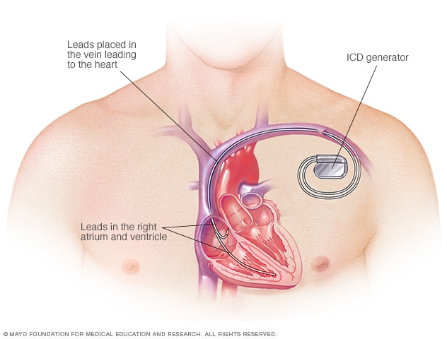
Implantable cardioverter-defibrillator (ICD)
An ICD controls the heartbeat by delivering shocks to the heart when the device detects an irregular heartbeat.
Subcutaneous implantable cardioverter-defibrillator (S-ICD)
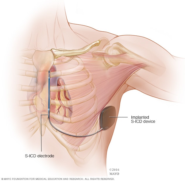
Subcutaneous implantable cardioverter-defibrillator (S-ICD)
A subcutaneous implantable cardioverter-defibrillator (S-ICD) is a less invasive alternative to a traditional ICD. The S-ICD device is placed under the skin at the side of the chest below the armpit. It connects it to a sensor that runs along the breastbone.
A surgery or procedure may be needed to control or prevent episodes of tachycardia.
- Cardioversion. This treatment is generally done when emergency care is needed for a long-lasting episode of ventricular tachycardia. Cardioversion uses quick, low-energy shocks to reset the heart rhythm. It's also possible to do cardioversion with medicines. A shock also can be delivered to the heart using an automated external defibrillator (AED).
- Catheter ablation. In this treatment, the doctor places one or more catheters into blood vessels to the heart. Sensors at the catheter tips use heat or cold energy to create tiny scars in your heart. The scars block irregular heart signals and restore the heartbeat. Catheter ablation also may be done to diagnose irregular heartbeats.
- Open-heart surgery. Some people with tachycardia need open-heart surgery to destroy the extra heart signaling pathway causing tachycardia. Such surgery is usually done when other treatments don't work or when surgery is needed to treat another heart condition.
Some people with tachycardia need a device to help control the heartbeat and reset the heart rhythm. Heart devices include:
- Implantable cardioverter-defibrillator (ICD). Your care team may suggest this device if you have a high risk of dangerously fast or irregular heartbeats in the lower heart chambers An ICD is placed under the skin near the collarbone. It continuously checks the heart rhythm. If the device finds an irregular heartbeat, it sends a shock to reset the heart's rhythm.
- Pacemaker. If slow heartbeats don't have a cause that can be fixed, a pacemaker may be needed. A pacemaker is a small device that's placed in the chest to help control the heartbeat. When it finds an irregular heartbeat, it sends an electrical signal that helps correct the heart's rhythm.
More Information
More Information
Clinical trials
Explore Mayo Clinic studies testing new treatments, interventions and tests as a means to prevent, detect, treat or manage this condition.
Coping and support
Make plans to manage an episode of a fast heartbeat. Doing so may help you feel calmer and more in control when one occurs. Talk to your care team about:
- How to check your heart rate and what rate is best for you.
- When to call your healthcare team.
- When to get emergency care.
Preparing for your appointment
If you have tachycardia, you may be sent to a doctor trained in heart diseases. This type of care professional is called a cardiologist.
If you have ventricular tachycardia, you may not have time to prepare. You may be treated at a hospital or emergency medical center.
If possible, bring someone with you who can provide support and help you remember any new information. Because there may be a lot to discuss, it may help to prepare a list of questions.
Make a list of:
- Your symptoms, including any that may seem unrelated to your heart.
- Important personal information, including any major stresses or recent life changes.
- Medicines that you take, including vitamins, supplements and those bought without a prescription. Include the dosages.
- Questions to ask your healthcare team.
List your questions from most important to least important in case time runs out. Basic questions include:
- What is likely causing my fast heart rate?
- What tests do I need?
- How do we check my heart?
- What's the most appropriate treatment?
- What are the risks and complications of ventricular tachycardia?
- How often do I need health checkups?
- I have other health conditions. How do I manage them together?
- Do I need to limit or avoid any activities?
- Are there any brochures or other printed material that I can take home with me? What websites do you recommend visiting?
Don't hesitate to ask additional questions.
What to expect from your doctor
When your heartbeat is controlled, your healthcare team is likely to ask you several questions. Your care team may ask:
- When did the symptoms start?
- How often do you have episodes of a fast heartbeat?
- How long do the episodes last?
- Does anything, such as exercise, stress or caffeine, seem to trigger or worsen episodes?
- Does anyone in your family have heart disease or a history of irregular heartbeats, called arrhythmias?
- Has anyone in your family had sudden cardiac death or died suddenly?
- Do you smoke?
- How much alcohol or caffeine do you use?
- Do you use substances such as cocaine or methamphetamines?
- Do you have high blood pressure, high cholesterol, or other heart or blood vessel conditions?
- What medicines do you take for these conditions, and do you take them as prescribed?