نظرة عامة
التليف الرئوي
التليف الرئوي
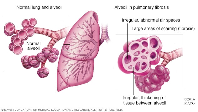
التليف الرئوي هو تندب وتيبس الأنسجة الموجودة حول الأكياس الهوائية (الحويصلات) في الرئتين وبينها، كما هو موضح على اليمين. وتظهر على اليسار رئة سليمة بها حويصلات سليمة.
التليف الرئوي مرض يصيب الرئة نتيجة تضرر نسيجها وتندّبه. يمنع هذا النسيج السميك والصلب الرئتين من العمل بشكل سليم. وتتفاقم حالة التليف الرئوي مع مرور الوقت. قد يبقى بعض المرضى في حالة مستقرة مدة طويلة، بينما تتدهور حالة آخرين بشكل أسرع. ويزداد ضيق النفس مع تفاقم الحالة.
يمكن أن ينتج التليف الرئوي عن عدد من العوامل. في كثير من الأحيان، لا يتمكن الأطباء وغيرهم من اختصاصيي الرعاية الصحية من تحديد سبب المشكلة. في حال عدم التوصُّل إلى السبب، تُسمَّى الحالة التليف الرئوي مجهول السبب.
يحدث التليف الرئوي مجهول السبب عادةً لدى البالغين الأكبر سنًا ومَن هم في منتصف العمر. يُشخَّص التليف الرئوي أحيانًا عند الأطفال والرُضَّع، لكن هذا ليس شائعًا.
لا يمكن ترميم الرئتَين المتضررتَين بسبب التليف الرئوي. يمكن أن تساعد الأدوية والعلاجات أحيانًا على إبطاء معدل التليُّف وتخفيف الأعراض وتحسين جودة الحياة. قد تكون زراعة الرئتَين خيارًا مناسبًا لبعض المرضى.
المنتجات والخدمات
الأعراض
يمكن أن تشمل أعراض التليف الرئوي ما يأتي:
- ضيق النفس.
- السعال الجاف.
- التعب الشديد.
- فقدان الوزن بغير قصد.
- وَجَع العضلات والمفاصل.
- زيادة عرض واستدارة أطراف أصابع اليدين والقدمين، ما يُسمى تعجّر الأظافر.
يمكن أن تختلف سرعة تفاقم التليف الرئوي بمرور الوقت ومدى شدة الأعراض بشكل كبير من شخص إلى آخر. يُصاب بعض الأشخاص بمرض شديد بسرعة. ويُصاب البعض الآخر بأعراض متوسِّطة تزداد سوءًا ببطء، على مدار شهور إلى سنوات.
عند تفاقم الأعراض بشكل مفاجئ
يمكن أن يزداد سوء ضيق النفس سوءًا على نحو مفاجئ لفترة تستمر من أيام إلى أسابيع لدى الأشخاص المصابين بالتليف الرئوي، وخصوصًا التليف الرئوي مجهول السبب. وتُسمى هذه الحالة التفاقم الحاد. وقد تكون مهددة للحياة. وقد يكون سبب التفاقم الحاد حالة أو مرضًا آخر، مثل عدوى الرئة. لكن عادةً ما يكون السبب مجهولاً.
الحالات التي تستلزم زيارة الطبيب
إذا كانت لديك أعراض التليف الرئوي، فتواصل سريعًا مع الطبيب أو اختصاصي رعاية صحية آخر. وإذا كانت الأعراض تزداد سوءًا، وخصوصًا إذا كان ذلك بسرعة، فتواصل مع فريق الرعاية الصحية فورًا.
الأسباب
التليف الرئوي تندّبٌ وثخانةٌ في الأنسجة المحيطة بالأكياس الهوائية، التي تُسمَّى الحويصلات الهوائية، والفاصلة بينها في الرئتين. وتُصعِّب هذه التغيرات انتقال الأكسجين إلى مجرى الدم.
قد يحدث تضرر الرئتين الذي ينتج عنه التليف الرئوي بسبب عوامل مختلفة. ومن أمثلتها التعرض لبعض المواد السامة مدة طويلة، والعلاج الإشعاعي، وتناوُل بعض الأدوية، والإصابة بحالات مرضية معينة. وفي بعض الحالات، يكون سبب الإصابة بالتليف الرئوي مجهولاً.
طبيعة العمل وبيئة العمل
قد يكمن السبب الرئيسي أو الجزئي للإصابة بالتليف الرئوي في نوع العمل الذي تؤديه والمكان الذي تعمل أو تعيش فيه. فالتعرض المستمر أو المتكرر للمواد السامة أو الملوِّثة، أي المواد التي تُفسد جودة الماء أو الهواء أو التربة، يمكن أن يضر الرئتين، خصوصًا في حال عدم ارتداء معدات واقية. ومن أمثلة ذلك:
- غبار السيليكا.
- الألياف الأسبستية.
- غبار المعادن الثقيلة.
- نشارة الخشب وغبار الفحم والحبوب.
- العفن.
- روث الطيور والحيوانات.
العلاجات الإشعاعية
تَظهر مؤشرات تضرر الرئة على بعض الأشخاص الذين يَتلقون علاجًا إشعاعيًّا على الصدر، مثل علاج سرطان الرئة أو الثدي، بعد العلاج بأشهر وأحيانًا بسنوات. وقد تعتمد مدى خطورة التضرر على:
- حجم جزء الرئة الذي تعرض للإشعاع.
- إجمالي كمية الإشعاع المُستخدمة.
- ما إذا كان استُخدِم العلاج الكيميائي أيضًا.
- ما إذا كان الشخص مصابًا بمرض كامن في الرئة.
الأدوية
قد تسبب أدوية كثيرة تضررَ الرئتين. ومن أمثلتها ما يأتي:
- العلاج الكيميائي. الأدوية المعدَّة لقتل الخلايا السرطانية، مثل ميثوتريكسات (Trexall، وOtrexup، وغيرهما) وبليوميسين وسيكلوفُسفاميد (Cytoxan)، يمكن أن تُتلف أنسجة الرئة.
- أدوية القلب. بعض الأدوية المستخدمة في علاج اضطراب نبض القلب، مثل الأميودارون (Nexterone، وPacerone)، يمكن أن تَضر أنسجة الرئة.
- بعض المضادات الحيوية. المضادات الحيوية مثل نتروفورانْتُوين (Macrobid، وMacrodantin) أو إيثامبيوتول (Myambutol) يمكن أن تُسبب تضررَ الرئة.
- الأدوية المضادة للالتهاب. بعض مضادات الالتهاب مثل ريتوكسيماب (Rituxan) أو سلفاسالازين (Azulfidine) يمكن أن تُسبب تضررَ الرئة.
الحالات الطبية
يُؤدي عدد من الحالات الصحية لتلف الرئة مثل:
- التهاب الجلد والعضل، وهو مرض التهابي يتميز بضعف العضلات والطفح الجلدي.
- الذئبة، وهو مرض يهاجم فيه جهاز المناعة في الجسم خلاياه وأعضاءه.
- مرض النسيج الضام المختلط، والذي يتميز بأعراض مختلطة لعدة اضطرابات، مثل الذئبة وتصلب الجلد والتهاب العضلات.
- التهاب الرئة، وهو عَدوى تؤدي إلى التهاب الأكياس الهوائية في إحدى الرئتين أو كلتيهما.
- التهاب العضلات، وهو مرض التهابي يُسبب ضعف العضلات على كلا جانبي الجسم.
- التهاب المفاصل الروماتويدي، وهو مرض التهابي يؤثر في المفاصل وأجهزة الجسم الأخرى.
- الساركويد، وهو مرض التهابي يؤثر في الرئتين والعُقَد اللمفية غالبًا.
- تصلب الجلد، وهو مجموعة من الأمراض النادرة التي تتضمن شد الجلد وتصلبه وكذلك مشكلات داخل الجسم.
التليُّف الرئوي مجهول السبب
تُسبب العديد من المواد والحالات الصحية التليف الرئوي. ومع ذلك، لا يُعرف سببها في العديد من الأشخاص. ولكن يمكن أن تكون عوامل الخطورة مثل التدخين أو التعرض لتلوث الهواء مرتبطة بهذه الحالة المرضية، حتى إذا تعذَّر تأكيد السبب. يُسمى التليف الرئوي من دون سبب واضح بالتليف الرئوي مجهول السبب.
قد يُصاب العديد من الأشخاص المصابين بالتليف الرئوي مجهول السبب أيضًا بداء الارتجاع المَعِدي المريئي، والمعروف أيضًا بـ GERD. وتحدث هذا الحالة عندما يتدفق الحمض من المعدة إلى المريء مرة أخرى. قد يكون داء الارتجاع المَعِدي المريئي من عوامل الخطورة المرتبطة بالتليف الرئوي مجهول السبب أو يُسبب تفاقم الحالة المرضية بشكلٍ أسرع. لكن يجب إجراء مزيد من الدراسات.
عوامل الخطر
يمكن أن يُصاب الأطفال والرضع بالتليف الرئوي، لكن ذلك ليس شائعًا. وتزداد احتمالية الإصابة بالتليف الرئوي مجهول السبب لدى الأشخاص في منتصف العمر والبالغون الأكبر سنًا. ويمكن أن يُصاب الشباب بأنواع أخرى من التليف الرئوي، مثل التي يسببها مرض النسيج الضام.
تشمل العوامل التي يمكن أن تزيد خطر الإصابة بالتليف الرئوي ما يأتي:
- التدخين. إذا كنت تدخن حاليًا أو كنت تدخن من قبل، فإنك عرضة بشكل أكبر لخطر الإصابة بالتليف الرئوي من الأشخاص الذين لم يدخنوا قط. كما يزداد خطر الإصابة به لدى الأشخاص المصابين بالنُّفاخ الرئوي.
- أنواع مِهن معينة. أنت معرض بشكل أكبر لخطر الإصابة بالتليف الرئوي إذا كنت تعمل في مجال التعدين أو الزراعة أو البناء. كما يزداد خطر الإصابة به في حال التعرض المستمر أو المتكرر للمواد الملوِّثة المعروفة بأنها تضر بالرئتين.
- علاجات السرطان. يمكن أن يزيد تلقِّي العلاجات الإشعاعية على الصدر أو استخدام بعض أدوية العلاج الكيميائي خطرَ الإصابة بالتليف الرئوي.
- الخصائص الوراثية. تسري بعض أنواع التليف الرئوي في العائلة، لذلك قد يكون للجينات دور في الإصابة بالمرض.
المضاعفات
قد تشمل مضاعفات التليف الرئوي ما يأتي:
- ارتفاع ضغط الدم في الرئتين. يُسمى بارتفاع ضغط الدم الرئوي، ويؤثر هذا النوع من ارتفاع ضغط الدم في شرايين الرئتين. وهي الشرايين الرئوية. قد تؤدي الشرايين المتصلبة والسميكة إلى إبطاء الدم أو منع تدفقه عبر الرئتين. ويؤدي ذلك إلى ارتفاع الضغط داخل الشرايين الرئوية وحجرة القلب اليُمنى السُفلية، التي تسمى البُطين الأيمن.
- فشل الجانب الأيمن من القلب. تحدث هذه الحالة الخطيرة عندما يتعين على حجرة القلب اليمنى ضخ الدم بقوة أكبر من المعتاد لنقل الدم عبر الشرايين الرئوية المسدودة جزئيًا.
- الفشل التنفسي. غالبًا ما تكون هذه المرحلة الأخيرة من مرض الرئة طويل المدى. وتحدث عند انخفاض مستويات الأكسجين في الدم بشكل خطر.
- سرطان الرئة. يزيد التليف الرئوي طويل المدى خطر الإصابة بسرطان الرئة.
- مشكلات الرئة الأخرى. مع تفاقم التليف الرئوي بمرور الوقت، قد يؤدي ذلك إلى مشكلات خطيرة مثل الجلطات الدموية في الرئتين أو انخماص الرئة أو حالات عَدوى الرئة.