May 21, 2021
Although rare, moyamoya disease can have devastating consequences, triggering strokes and seizures in children and adults. Mayo Clinic's new imaging protocol for managing the disease includes various MRI sequences that can be completed in the same session to safely and efficiently determine whether a patient needs surgical intervention.
"Ideally, we want to diagnose moyamoya disease early and select patients who are at risk of future strokes and require surgery versus patients who can be managed with medical therapy alone," says Giuseppe Lanzino, M.D., a cerebrovascular neurosurgeon at Mayo Clinic in Rochester, Minnesota. "With our new protocol, we can get the information we need with a single MRI. In the past, different imaging modalities were required."
Detailed moyamoya imaging protocol
Detailed moyamoya imaging protocol
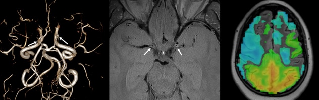
Left. 3D MR angiography of a young adult with moyamoya disease demonstrates tapered narrowing of both internal carotid termini and occlusion directly at the termini (white arrows). Center. High-resolution vessel wall imaging demonstrates negative remodeling of the tapered portions of the internal carotid arteries with mild diffuse circumferential wall enhancement (white arrows). Right. Cerebrovascular reactivity map from a 20-second breath hold blood oxygen level dependent (BOLD) exam demonstrates areas of reduced cerebrovascularity reactivity throughout most of the visualized middle cerebral artery and anterior cerebral artery territories bilaterally (blue).
The approaches used for imaging in moyamoya disease vary greatly, but generally involve multiple patient visits and often include nuclear medicine testing. Mayo Clinic's protocol instead combines several MRI components, including vessel wall imaging and blood oxygenation level-dependent (BOLD) functional MRI.
"The imaging protocol that we've established provides a deep assessment of the brain and the affected blood vessels without ionizing radiation. That's important because moyamoya disease typically affects younger people," says Vance T. Lehman, M.D., a neuroradiologist at Mayo Clinic's Rochester campus. "We do identical imaging tests at follow-up visits for consistency in tracking disease progression."
Moyamoya disease is a progressive disorder characterized by blockage in the distal internal carotid artery and adjacent blood vessels. The name moyamoya, which means "puff of smoke" in Japanese, describes the appearance of the tangle of tiny blood vessels formed to compensate for the blockage. Moyamoya disease can occur at any age, but symptoms are most common in children ages 5 to 10 and adults ages 30 to 50.
Under the leadership of Fredric B. Meyer, M.D., enterprise chair of Neurologic Surgery at the Rochester campus, Mayo Clinic has developed extensive experience managing moyamoya disease. In a three-part series published in 2020 in Contemporary Neurosurgery, Mayo Clinic neurological specialists outlined patient presentations of moyamoya disease as well as optimal current approaches to diagnosis and treatment.
"As a high-volume center, Mayo Clinic can dedicate a subspecialized team to the diagnosis, treatment and research of moyomoya disease," Dr. Lanzino says. "The team is able to give patients the follow-up care they need over time."
Cutting-edge MRI
Diagnosis is challenging, as moyamoya disease has many mimics. Some patients have moyamoya syndrome, defined as moyamoya caused by an underlying condition such as Down syndrome or neurofibromatosis. Precise diagnosis is crucial because the treatment pathways for moyamoya disease differ from the approaches to other conditions.
High-resolution MRI of the cerebral blood vessel walls can reveal patterns that are distinctive to moyamoya disease and its mimics. "Vessel wall imaging allows us to evaluate cerebral blood vessel walls with submillimeter resolution," Dr. Lehman says. "That helps us to differentiate idiopathic moyamoya disease from other conditions."
Changes in cerebral hemodynamics are another key factor. BOLD MRI helps identify individuals with impaired cerebrovascular reserve when under physiological stress. "These patients can become symptomatic and may be at risk of stroke," Dr. Lanzino says.
Patients who are asymptomatic and not at immediate risk of infarction might be closely monitored or managed medically with blood thinners and correction of risk factors. When needed, surgery involves direct or indirect revascularization, or combined direct and indirect revascularization.
"Treatment decisions are complex and involve many factors," Dr. Lanzino says. "An experienced team is key to providing optimal care individualized to the patient."
Cerebrovascular headaches
Mayo Clinic's approach to moyamoya disease includes managing patients' cerebrovascular headaches. "Due to diminished blood flow in the brain, patients with moyamoya disease often experience headaches that are very difficult to treat," Dr. Lanzino says. The moyamoya treatment team at Mayo Clinic's Rochester campus now includes Chia-Chun Chiang, M.D., a neurologist with subspecialty training in headache and cerebrovascular disease.
Individuals with moyamoya disease require lifelong care. Mayo Clinic performs regular follow-up clinical visits and imaging to determine whether surgery has been successful and patients' vascular bypasses have matured, and to monitor patients who have been treated with medical therapy alone.
Anecdotal evidence suggests that moyamoya disease, originally described in East Asian populations, might be increasing in prevalence among Western populations. "It's difficult to know if there is a true increase in the disease incidence or if we have a lower threshold for suspecting moyamoya disease and our tests are finding it," Dr. Lanzino says. "But there is no question that over time, we are seeing more patients with moyamoya disease."
For more information
Larson AS, et al. Contemporary management of moyamoya disease: Part I — Background and clinical presentation. Contemporary Neurosurgery. 2020;42(6):1
Larson AS, et al. Contemporary management of moyamoya disease: Part II — Imaging features and grading systems. Contemporary Neurosurgery. 2020;42(7):1.
Larson AS, et al. Contemporary management of moyamoya disease: Part III — Revascularization techniques. Contemporary Current Neurosurgery. 2020;42(8):1.
Refer a patient to Mayo Clinic.