Overview
Positron emission tomography (PET)

Positron emission tomography (PET)
During a positron emission tomography (PET) scan, a healthcare professional injects a small amount of a radioactive substance called a tracer into a vein. You then lie on a narrow table that moves into a large, ring-shaped machine. The scanner makes pictures that show how cells and tissues take up the tracer. A PET scan can help diagnose conditions such as cancer, heart disease and brain conditions. The test also may be done to help plan treatment.
A positron emission tomography (PET) scan is an imaging test that shows how your body's tissues and organs are working. The test uses a small amount of a radioactive substance called a tracer. Depending on the reason for the test, the tracer may show how cells use sugar, how blood flows, or how some proteins or other substances attach to cells. PET scans can help find changes linked to diseases such as cancer or brain conditions such as Alzheimer’s disease.
A PET scan often can find changes earlier than other imaging tests, such as a CT scan or an MRI scan.
A PET scan is usually done with a CT scan in a single test called a PET-CT scan. The PET scan shows how the body is working. The CT scan shows the structure of the body, such as its size and shape. PET-CT gives more complete and more accurate information than either test does alone. If a PET scan is done with an MRI, it's called PET-MRI.
Positron emission tomography (PET) is not the same as single-photon emission computerized tomography (SPECT). A SPECT scan uses different tracers and gives less detailed images than a PET scan does.
Types
All PET scans use a small amount of radioactive substance called a tracer. Sometimes the type of PET scan is named for the tracer used. Examples are:
- FDG PET scan. This is the most common type of PET scan. FDG, also called fluorodeoxyglucose, is a type of sugar. An FDG PET scan shows how cells use sugar. Cancer cells typically use more sugar, so this test can help find or stage some cancers.
- Gallium PET scan. This type of PET scan uses a tracer such as gallium-68 to look at some types of tumors or infections. A DOTATATE PET scan is a type of this scan that may be used for some tumors.
- Rubidium PET scan. This PET scan often is done to check blood flow to the heart.
- Other PET tracers. Some PET scans use special tracers for some conditions. For example, an amyloid PET scan may be used for brain conditions such as Alzheimer's disease. A PET scan using a tracer called fluciclovine (Axumin) may sometimes be used to find prostate cancer.
Specific types of PET scans include:
Products & Services
Why it's done
A PET scan is done to see how the body's cells and tissues are working. The scan can look at many parts of the body, including the brain, lungs and heart.
Your healthcare professional may do a PET scan to:
- Find cancer or see how much it's spread.
- Plan or monitor treatment for cancer and other conditions.
- Diagnose brain conditions such as Alzheimer's disease.
- Look at blood flow in the heart to find heart disease.
Sometimes a whole-body PET scan is done to look for cancer in different parts of the body, such as when cancer has spread or come back. A whole-body PET scan usually goes from head to feet or from the skull to the thighs.
Information from a PET scan can help diagnose, watch or treat a condition. A healthcare professional must read a PET scan carefully. Some things, such as inflammation or noncancerous conditions, can look like cancer on a PET scan. Also, PET scans may be less accurate in people with diabetes. That's because blood sugar levels can affect how the tracer is taken up by cells.
PET scans for cancer
PET plus CT
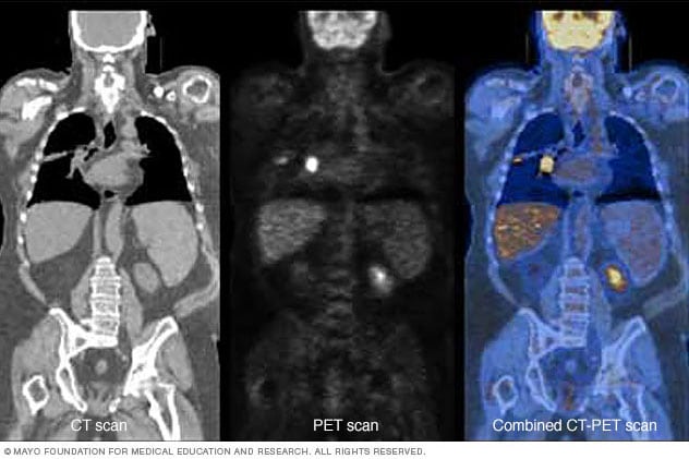
PET plus CT
Combining a PET scan with a CT scan can make the images easier for a healthcare professional to read. The image on the left shows a CT scan. The center image shows a PET scan. The image on the right shows a combined PET-CT scan. The bright area in the chest, seen most clearly on the PET and PET-CT images, shows lung cancer.
A PET scan is very helpful in finding cancer. It can be more accurate than other imaging tests alone. Before a PET scan, you get a small amount of a radioactive substance called a tracer. The most common tracer used for cancer scans is fluorodeoxyglucose (FDG), which is a form of sugar. The scan shows where the tracer collects in cells. Cancer cells show up as bright spots on PET scans because they usually use more sugar than do typical cells.
A PET scan may help:
- Find cancer.
- Show whether cancer has spread.
- Check whether a cancer treatment is working.
- See if cancer has come back.
A PET scan combined with a CT or an MRI scan can help find many types of cancer, such as:
- Brain cancer. A PET scan is not usually the first test used to check for brain cancer. It may be done if other imaging results are not clear. It also may be used to tell the difference between a brain tumor and treatment-related changes such as scar tissue.
- Breast cancer. If you have breast cancer, your healthcare team may use a PET scan to learn the stage. The stage shows how much cancer is in the body and if it has spread.
- Colorectal cancer. A PET scan can help find colorectal cancer that has come back and check to see whether it has spread. Colorectal cancer includes cancers of the colon and rectum.
- Head and neck cancer. A PET scan can help find head and neck cancer, including the main tumor and areas where it has spread.
- Lung cancer. A PET scan can help find lung cancer, show how far it has spread and guide treatment.
- Lymphoma. Lymphoma starts in the lymphatic system, which is part of the body's immune system. A PET scan can help find Hodgkin lymphoma and some non-Hodgkin lymphomas and check how well treatment is working. But some slower growing types of lymphoma may not show up as brightly on a PET scan and can be harder to find.
- Prostate cancer. A PET scan may be used if prostate cancer has come back or spread, especially when other test results are not clear. But some types of prostate cancer may not show up well on a standard PET scan because they do not use as much of the sugar tracer. A PSMA PET scan may be done to better show prostate cancer. This scan targets a protein found on prostate cancer cells.
PET-CT also may be done for these cancers:
- Cervical cancer. A PET scan can tell whether cervical cancer has spread to other parts of the body.
- Esophageal cancer. A healthcare professional may order a PET scan to learn the stage of cancer of the esophagus and help plan treatment.
- Melanoma skin cancer. If you have this type of skin cancer, your healthcare team may use a PET scan to see whether it has spread to other parts of the body.
- Pancreatic cancer. If you have pancreatic cancer, your healthcare team may use a PET scan to see whether it has spread to other parts of the body and to help plan treatment. It also may be used to check how well treatment is working.
- Thyroid cancer. A PET scan may be done when blood tests suggest some types of thyroid cancer have come back but other imaging tests do not clearly show where it is. It can help find cancer that has spread or returned.
PET scans for brain conditions
PET scan of the brain for Alzheimer's disease

PET scan of the brain for Alzheimer's disease
A PET scan can compare a typical brain (left) with one affected by Alzheimer's disease (right). Less red and more yellow, blue and green can show parts of the brain that use less energy (sugar) because of Alzheimer's disease.
A PET scan can be used to check some brain conditions such as tumors, Alzheimer's disease and seizures. Many PET scans show how active different parts of the brain are by checking how the brain uses sugar for energy. This can help find areas of the brain that are not working as expected.
Some PET scans use a tracer called an amyloid tracer. This type of scan can show amyloid plaques in the brain, which are linked to Alzheimer's disease.
A brain PET scan can help your healthcare professional:
- See patterns of brain activity linked to Alzheimer's disease or other types of dementia.
- Find where seizures start in the brain.
- Tell the difference between a brain tumor and scar tissue.
PET scans for heart disease
PET scan of the heart
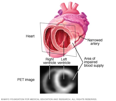
PET scan of the heart
This PET scan of the heart, also called a cardiac PET scan, shows a darker area that may mean less blood flow from a narrowed artery. The reduced blood flow is in one of the lower heart chambers, called the ventricles. Information from a heart PET scan can help your healthcare professional plan treatment.
When a PET scan is used to look at the heart, it is called a cardiac PET scan or heart PET scan. It can show areas of the heart that are not getting enough blood, often due to a narrowed or blocked artery. A PET scan also can show whether the heart muscle is still healthy or has been damaged.
The information from a heart PET scan helps you and your healthcare professional decide what to do next. For example, a PET scan may show whether a treatment such as angioplasty or coronary artery bypass surgery could improve blood flow. It also can show how a heart attack has affected your heart.
A PET scan of the heart may be done in different ways depending on what your healthcare team needs to learn.
- Myocardial perfusion PET scan. This test is sometimes part of a nuclear stress test. It shows how well blood is moving through the heart muscle. It can help show parts of the heart that are not getting enough blood.
- PET myocardial viability scan. Also called a cardiac PET viability scan, this test shows whether heart muscle is still alive and may get stronger after treatment. Your healthcare team can use the results to see whether improving blood flow could help your heart work better.
- Cardiac PET metabolism scan. Also called an FDG cardiac PET scan, this test shows how the heart muscle uses sugar for energy. Areas that use more sugar appear brighter, while areas that use less appear darker. Darker areas may mean the heart muscle is damaged. But areas of low sugar use do not always mean damage. Your healthcare professional compares them with blood flow to see whether the heart muscle is scarred or still alive.
PET scans for inflammation
PET scans can show areas of inflammation or infection. Many PET scans use a tracer that acts like sugar to show how active cells are. Areas of inflammation or infection may look brighter because these cells use more sugar. However, PET scans can't always tell the difference between inflammation, infection and cancer.
A healthcare professional may do a PET scan to look for conditions such as:
- Appendicitis.
- Inflammation of the gallbladder, called cholecystitis.
- Inflammatory bowel disease.
- A bone infection called osteomyelitis.
- Inflammation of the pancreas, called pancreatitis.
- Growth of tiny clumps of immune cells in the body, called sarcoidosis.
- Swelling of the thyroid gland, called thyroiditis.
- A lung infection called tuberculosis.
- Swelling of blood vessels, called vasculitis.
Sometimes, a PET scan is done to find inflammation after a biopsy or surgery. This helps your healthcare professional learn whether an area is healing as expected or there may be infection or another condition.
More Information
Risks
A PET scan uses a small amount of radiation, which is measured in millisieverts (mSv). A PET scan exposes you to about 10 mSv of radiation. A PET scan done with CT exposes you to about 20 mSv. These levels of radiation are considered low. Your healthcare team takes steps to keep your exposure as low as possible.
For most people, the radiation from a PET scan is considered safe. Even if you need more than one scan, the total radiation exposure is similar to that of other imaging tests, such as CT scans.
Some people may worry that PET scans cause cancer. Any exposure to radiation may carry a very small risk of cancer over time. But the risk from a PET scan is low, and the benefits of the test often outweigh this risk. Your healthcare professional can help you understand your personal risk.
Other possible PET scan risks include:
- If you're pregnant, radiation may reach the unborn baby, also called a fetus.
- Radiation may pass through breast milk to a baby.
- An allergic reaction, although this is rare.
Talk with your healthcare professional about the benefits and risks of a PET scan.
How you prepare
Some things may affect PET scan results. Your healthcare team tells you how to prepare for a PET scan. Follow these tips to get ready.
- Exercise. Do not do heavy exercise for at least a day before the scan. Exercise can affect how your body uses sugar and may change the results.
- Food and drinks. Your healthcare team usually tells you not to eat or drink for about 4 to 6 hours before the scan. This is called fasting. Drink only water unless your healthcare team says other liquids are OK. Drinks such as coffee or alcohol may affect how your body uses the tracer and may change the results. You can likely eat and drink as usual after the scan unless your healthcare team tells you otherwise.
- Clothing. Wear comfortable, loose-fitting clothes to the test.
- Medicines. Tell your healthcare professional about all medicines, vitamins and supplements you take. You may be given special instructions, especially if you have diabetes. Diabetes can affect how cells use the tracer given during the test.
Follow your healthcare team's instructions to help make sure your PET scan results are accurate.
Tell your healthcare professional if:
- You are pregnant or think you might be pregnant.
- You are breastfeeding.
- You have had a reaction to the tracer used for the PET scan.
- You feel sick or have any medical conditions.
If being in a closed space makes you anxious, or you're worried about the PET scan, tell your healthcare professional when you schedule the test. You may get medicine to help you relax during the scan.
What you can expect
A PET scan is done in a hospital or medical center. The scan may be done with a CT or an MRI scan to give more detail. The scanner is a large machine shaped like a ring.
Before
Before the PET scan starts, your healthcare team may ask you to change into a gown. You also may be asked to take off any metal objects such as jewelry.
During
A PET scan is done in two parts. First, a member of your healthcare team places a small amount of radioactive tracer into a vein in your arm or hand. The most common tracer is called fluorodeoxyglucose (FDG). Different PET scans may use different tracers depending on why you are having the test and the area being studied. You may briefly feel cold as it goes into your body. You then rest quietly for about 30 to 60 minutes while your body soaks up the tracer.
After the tracer has had time to travel through your body, you lie on a narrow, padded table that slides into a scanner. The scanner may make buzzing or clicking sounds. You need to lie very still so the images are clear. The scan itself usually takes about 30 minutes but may take longer depending on the reason for your test.
The scan shows how cells soak up the tracer. The FDG tracer shows how your cells use sugar for energy. Cells that are more active use more sugar and take in more FDG. This is called increased uptake.
The full test usually takes about 2 hours. Most people go home the same day.
After the procedure
Most people go home the same day as their PET scans. Some people may need to stay longer depending on their condition or why the test is done.
After the test, you can go back to your usual activities unless your healthcare team tells you not to. If you get medicine to help you relax during the test, you need someone to drive you home. Drink plenty of water to help remove the tracer from your body.
Results
A healthcare specialist called a radiologist reads your PET scan and sends results to your healthcare team. A written report may be available in a few days but the time depends on your medical center. Your healthcare professional can use PET scan results to help diagnose, watch or treat your condition.
PET scan results may include this information:
- Increased uptake. This means more of the PET scan tracer is seen in an area. Increased uptake on a PET scan may be a sign of cancer, inflammation or infection. But it depends on the reason for the test.
- Hypermetabolic activity. This means the cells in some areas are more active and use more energy. These areas often show increased uptake on a PET scan.
- Standardized uptake value (SUV). SUV is a measurement of how much tracer your cells and tissues take up. The value varies from person to person and by the area being tested. There is no single SUV value or standard range that applies to everyone. A higher SUV often means increased uptake. Your healthcare team may use your SUV to track changes over time.
False-positive result
A PET scan is not always exact. Sometimes it may show something that looks like cancer when it is not. This is called a false-positive. An example of a false-positive on a PET scan is when areas of inflammation or infection take in more tracer and look like cancer on the scan.
False-negative result
A PET scan may miss very small cancers. Also, some types of cancer, such as prostate cancer, may not show up as clearly because they do not use as much sugar. When this happens, the cancer is said to have low FDG uptake.
Your healthcare professional uses PET scan results along with other tests to make a diagnosis.
After a PET scan, your healthcare team may need to remove some tissue for examination to confirm a diagnosis. This is called a biopsy. A PET scan may help guide your healthcare professional about where to take a biopsy, but it does not replace a biopsy.
Clinical trials
Explore Mayo Clinic studies of tests and procedures to help prevent, detect, treat or manage conditions.