April 27, 2018
Mayo Clinic is integrating several sophisticated techniques to provide an advanced approach to endoscopic skull base surgery. These innovations, available at all three Mayo Clinic campuses, enhance the precision of complex skull base surgeries.
"We are bringing together many high-end pieces to provide integrative techniques that don't happen in a lot of other places," says Bernard R. Bendok, M.D., chair of Neurosurgery at Mayo Clinic in Phoenix/Scottsdale, Arizona. "It's an intense area of focus for us, and we are getting referrals from around the world."
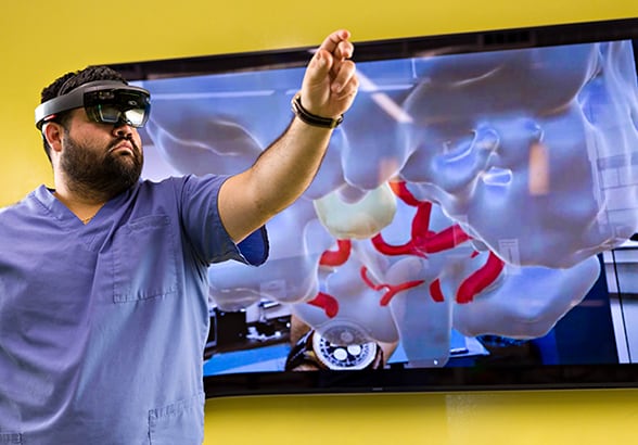 Holographic version of a brain before surgery
Mr. Welz walks through a holographic version of a brain, complete with arteries and tumor, to allow for visualization of the space before surgery.
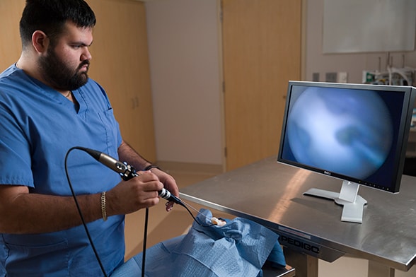 3-D printed anatomy demonstration
Matthew Welz, a research technologist at Mayo Clinic in Phoenix/Scottsdale, Arizona, demonstrates 3-D printed anatomy that is used to practice surgical removal of skull base tumors.
These innovations, used in surgeries to treat pituitary tumors, meningiomas and clival tumors, include:
- Image guidance and intraoperative CT
- 3-D endoscopy
- Simulations that integrate endoscopic surgery and proton beam therapy
- 3-D models and holography for pre-surgical planning
Mayo Clinic's integrated, multidisciplinary practice means that an ENT surgeon works alongside the neurosurgeon throughout treatment. "That's one of our biggest advantages," says Devyani Lal, M.D., a consultant in Otorhinolaryngology specializing in rhinology-skull base surgery at Mayo Clinic's campus in Arizona. "Skull base pathology requires combined ENT and neurological expertise. Our integrated approach maximizes our ability to remove tumors completely."
High-end technology in many hands
Intraoperative CT provides enhanced visualization to maximize tumor resection. "We're able to resect a lot of the tumor with less risk to the cranial nerves. We also get a better view of the cavernous sinus, so we can work more safely," Dr. Bendok says. "Craniotomy is avoided, and we're able to remove skull base tumors less invasively."
Angled endoscopes further enhance visualization and surgical precision. "We can look around corners and find a tumor even if it's hidden behind the carotid artery or in the sphenoidal sinus or up high in the skull base," Dr. Lal says. "The collaboration between ENT and neurosurgery is a very dynamic process. I work with an endoscope every day of my professional life. During skull base surgeries, I often suggest looking for tumor in certain places. Four hands are better than two, and two brains are better than one."
Endoscopic techniques are further enhanced via computer simulations that map how that surgical approach can be integrated with proton beam treatment. "That is a fabulous hybrid therapy for chordomas," Dr. Bendok says. "We use computer simulation to get a volumetric rendering of the tumor. Then, depending on how much tumor we remove, we can determine the dose of radiation that's needed. Simulations allow us to do the surgery before the surgery."
The latest innovation in pre-surgical planning involves augmented reality and holography. "We're incorporating the patient's anatomy into holography to create holograms of pituitary tumors," Dr. Bendok says. "We can walk through the patient's anatomy before surgery."
Lower risk, higher function
At Mayo Clinic, neurosurgeons and ENT surgeons meet weekly to discuss skull base cases. "Because of this combined effort, a multidisciplinary plan is fashioned that is individualized to the patient," Dr. Bendok says. "We use nasoseptal flaps and other ancillary flaps for skull base repair, resulting in negligible rates of postoperative cerebral spinal fluid leak. We've also been doing preoperative embolization and preoperative balloon test occlusion, when necessary, to reduce the risk of stroke from surgery." After skull base surgery, Mayo Clinic patients have specialized neurointensive care.
In addition to minimizing risk, Mayo Clinic strives to enhance patient recovery and quality of life. "We don't do packing of the nose routinely after skull base surgery. As a result, patients are much more comfortable," Dr. Lal says. "We also focus on functional outcomes. For many of our patients, we are able to restore vision and preserve olfactory function. For secretory pituitary tumors, we shoot for endocrinological cure.
"Our reconstructive surgeons are also involved in resection of large sinonasal and skull base malignancies with intracranial extension, such that needed cancer resection is never compromised," she adds. "This has helped with our local control and survival rates. No matter how large the resection, our reconstructive surgeons are able to fashion regional and free flaps to repair the defect for pleasing cosmetic and functional outcomes."
Mayo Clinic's approach can provide successful care even for people with complex disease. Dr. Lal cites a patient who came to Mayo Clinic's campus in Arizona with a meningioma pressing on her optic nerve.
"The tumor was in a very difficult location and causing visual compromise," Dr. Lal says. Reluctant to use radiation near the optic nerve, Dr. Lal and Dr. Bendok opted for surgical resection, approaching the tumor toward the midline.
"We had to be certain that the central retinal artery wasn't injured during tumor resection," Dr. Lal says. "Our neuroradiologist mapped out where the artery was, and then we did the surgery. We got all the tumor out, and she left the hospital the next day, with no nasal packing. The patient's visual loss and field deficit were restored on testing by our ophthalmologists. And although we worked through her nose for about eight hours, she smells and breathes beautifully."
"Providing this level of high expertise requires strong collaboration, not just with our neurosurgeons and ENT surgeons but also with our neurologists, neuroradiologists, endocrinologists and ophthalmologists," Dr. Bendok adds. "We bring that expertise to every one of our patients."