Overview
Double-outlet right ventricle (DORV) is a rare heart condition that is present at birth. A heart condition a child is born with is called a congenital heart defect. This is a change in the heart's structure that is present at birth.
The heart has two lower chambers, called ventricles. Two large arteries carry blood away from the heart: the aorta and the pulmonary artery. The aorta connects to the left ventricle and carries blood to the body. The pulmonary artery connects to the right ventricle and carries blood to the lungs. A wall separates the two ventricles. In babies with double-outlet right ventricle, the aorta and the pulmonary artery connect to the right lower heart chamber, either partially or completely.
Most babies with DORV also have an opening in the wall between the two ventricles. This is called a ventricular septal defect (VSD).
DORV can look different from one child to another. Blood flow and the type of repair needed can be affected by:
- The location of the VSD.
- The position of the aorta and the pulmonary artery.
- Narrowing in the pathways between the ventricles and the large arteries.
Most babies with DORV need surgery. The timing of surgery depends on your baby's heart structure, symptoms and blood flow. The healthcare team determines the type of treatment based on your child's heart structure.
With surgical treatment, most children have good long-term survival, although some may need additional heart procedures or lifelong follow-up.
Double-outlet right ventricle
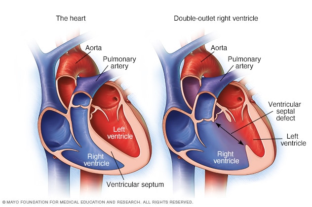
Double-outlet right ventricle
In double-outlet right ventricle, the lower right heart chamber, called the right ventricle, has two openings for blood to exit instead of one. Some blood goes to the body instead of just the lungs. There also is a hole between the two lower heart chambers. The hole is called a ventricular septal defect. In a typical heart, shown on the left, the pulmonary artery connects to the right ventricle and the aorta connects to the left ventricle.
Types
There are several types of double-outlet right ventricle (DORV). The difference between these types is the location of the ventricular septal defect (VSD) and whether it is close to or far from the pulmonary artery and the aorta. The type of DORV determines which treatment may be right for your child.
Subaortic VSD
With subaortic VSD, the opening between the ventricles is close to the aorta. Because both arteries connect to the right ventricle in DORV, the left ventricle does not have its usual direct path to the aorta.
Subpulmonary VSD and Taussig-Bing type
With subpulmonary VSD, the opening between the ventricles is close to the pulmonary artery. Because of this, blood from the left ventricle tends to flow toward the pulmonary artery instead of the aorta. Taussig-Bing type is a form of DORV with this type of VSD.
Doubly committed VSD
With doubly committed VSD, the opening between the ventricles is close to both the aorta and the pulmonary artery. Because of its location, blood from the left ventricle can reach either artery.
Noncommitted VSD
With noncommitted VSD, also called remote VSD, the opening between the ventricles is farther from both the aorta and the pulmonary artery.
Fallot-type DORV
With Fallot-type (fa-LOW type) DORV, the VSD is usually close to the aorta or close to both the aorta and pulmonary artery. The pathway that carries blood from the right ventricle to the pulmonary artery may be narrowed. This narrowing lessens the amount of blood that can reach the lungs.
This is called Fallot-type DORV because it can resemble tetralogy of Fallot, which is another congenital heart defect that can lessen blood flow to the lungs.
Products & Services
Symptoms
Signs and symptoms of DORV depend on how much blood flows from the heart to the lungs and to the rest of the body. Symptoms also depend on how much oxygen is in the blood.
Symptoms may start soon after birth or become more noticeable during the first weeks or months of life.
Symptoms may include:
- Blue or gray skin, lips, or nail beds. This can happen when the blood has too little oxygen. The color change may be easier or harder to see depending on skin color.
- Fast breathing or trouble breathing.
- Not eating well.
- Slow weight gain.
- Low energy or unusual sleepiness.
When to see a doctor
If your baby has any of the symptoms of DORV, contact your healthcare professional right away.
Children with DORV also need regular checkups with a congenital heart doctor, called a cardiologist. This specialist treats heart conditions present at birth. These checkups are important even when your child feels well because DORV and the effects of earlier heart surgery can change over time.
When to seek emergency care
Seek emergency medical care if your baby:
- Has severe trouble breathing.
- Looks much more blue or gray than usual.
- Is very sleepy or is hard to wake.
Causes
DORV develops before birth while the heart and large arteries are forming. The exact cause is not known. Changes in genes or chromosomes that happen before birth may affect how the heart develops and may play a role in DORV.
Risk factors
There is no single known risk factor for DORV. A family history of congenital heart disease or a known genetic condition may increase the chance of a baby having a congenital heart disease, such as DORV.
Other conditions and DORV
DORV can resemble another congenital heart condition called transposition of the great arteries. In this condition, the two main arteries leaving the heart connect to the opposite ventricles rather than to their usual positions. The aorta comes from the right ventricle, and the pulmonary artery comes from the left ventricle.
Babies with DORV also may have other health conditions, such as:
- Heterotaxy. This is a congenital condition in which organs in the chest or abdomen are arranged differently from their usual positions.
- Hypoplastic left heart syndrome. This is a congenital heart condition in which the left side of the heart is severely underdeveloped. Some babies with DORV also have a very small left ventricle and other heart differences that can block blood flow through the left side of the heart.
Complications
Complications can happen before surgery to repair DORV, such as:
- Too little oxygen in the blood.
- Too much blood flow to the lungs.
- Damage to blood vessels in the lungs.
- Slow growth.
- Heart failure. Heart failure happens when the heart cannot pump enough blood to meet the body's needs.
Sept. 10, 2026