概述
During a pterional (ter-e-O-nul) craniotomy, the surgeon temporarily removes part of the skull on the side of the head, in front of and above the ear. This surgery also is called a frontotemporal craniotomy.
The surgery may be used to reach brain tumors, bleeding in the brain and blood clots. It also may be used to reach a bulging blood vessel in the brain, called a brain aneurysm, or a blood vessel that did not form properly, called a vascular malformation.
Pterional craniotomy
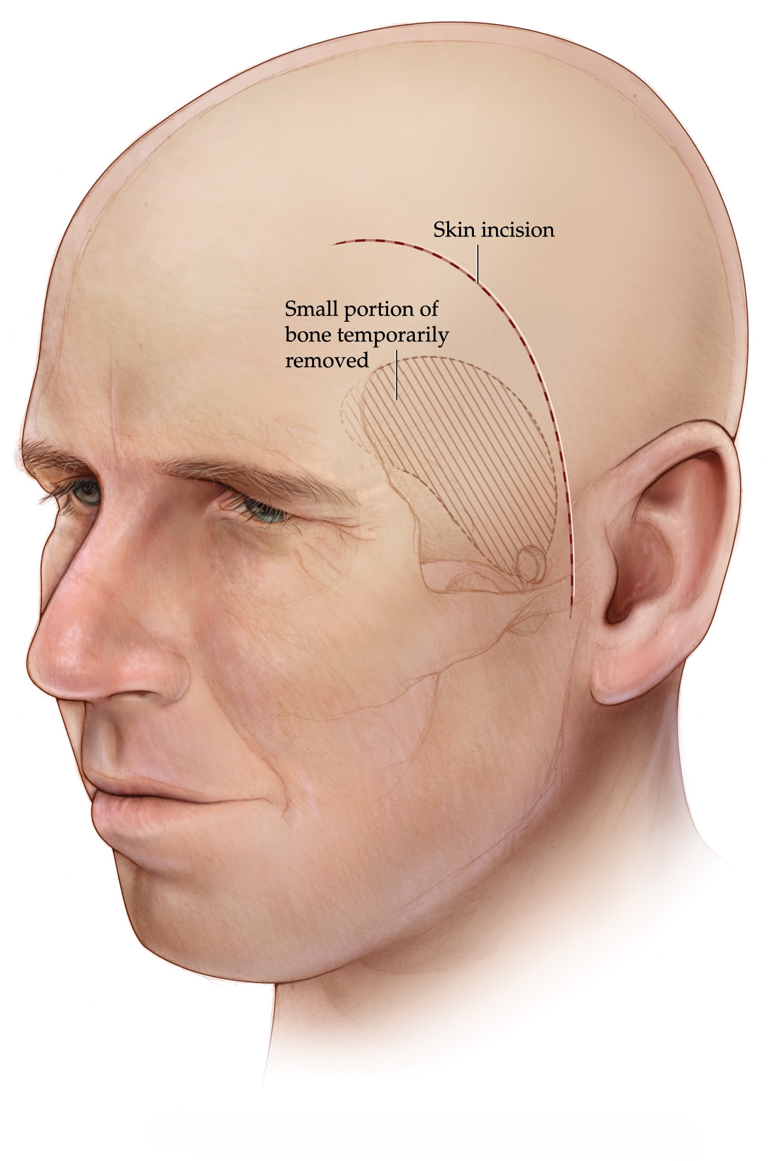
Pterional craniotomy
During a pterional craniotomy, a piece of skull is temporarily removed to expose the tough covering over the brain, called the dura mater, and the brain.
Types
A standard pterional craniotomy removes part of the skull on the side of the head in front of and above the ear. Variations of this surgery change the size or location of the opening so the surgeon can reach the area being treated.
- Pterional keyhole and minipterional craniotomies use smaller openings in the skull. A keyhole craniotomy removes bone near the outer edge of the eye and forehead. It can be used to reach brain aneurysms. A minipterional craniotomy removes bone near the pterion and behind the hairline. The pterion is the area in the temple where four skull bones meet. A minipterional craniotomy can be used to reach some tumors in or near the front and middle parts of the skull base.
- Extended and orbitozygomatic pterional craniotomies. These surgeries remove more bone to give the surgeon extra room or a wider angle to reach the area being treated. An extended pterional craniotomy removes more bone near the eye socket and the sphenoid wing. It can be used to reach large skull base tumors located in the front or middle part of the skull base. An orbitozygomatic craniotomy removes bone near the eye socket and cheekbone. It can be used to reach aneurysms and tumors in the front and middle parts of the skull base.
- Frontotemporal orbitozygomatic pterional craniotomy. This surgery combines a pterional craniotomy opening with removal of part of the eye socket and the cheekbone. It can be used to reach complex tumors deep in the skull base and around the pituitary gland.
A pterional craniotomy may be done on the left or right side of the head. The surgeon chooses the craniotomy type and side that give safe access to the area being treated while disturbing the brain and blood vessels as little as possible.
目的
Surgeons use a pterional craniotomy to reach some tumors and blood vessel conditions in the front and side areas of the skull base.
Tumors
A pterional craniotomy may be used to remove tumors in the front and middle areas of the skull base. Tumors that may be reached with a pterional craniotomy include:
- Meningiomas. A meningioma is a tumor that grows in the membranes that cover the brain, called the meninges. A pterional craniotomy may be used to reach some meningiomas near the front of the skull base and optic nerves.
- Pituitary tumors. A pituitary tumor is a growth in the pituitary gland at the base of the brain. A pterional craniotomy may be used to reach large tumors or tumors that involve nearby brain tissue or nerves.
- Craniopharyngiomas. A craniopharyngioma is a tumor that begins near the pituitary gland and can affect nearby structures in the brain. A pterional craniotomy may be used to reach some of these tumors, depending on where the tumor is located.
- Gliomas. A glioma is a tumor that begins in cells that support nerve cells. A pterional craniotomy may be used to reach gliomas in some areas of the frontal and temporal lobes.
Blood vessel conditions
A pterional craniotomy also may be used for some conditions that involve blood vessels in the brain.
- Brain aneurysm. A brain aneurysm is a bulge or ballooning in a blood vessel in the brain. A pterional craniotomy may be used to reach brain aneurysms that have or have not ruptured.
- Arteriovenous malformation (AVM). An AVM is a tangle of blood vessels in which blood flows directly from arteries to veins instead of through smaller blood vessels called capillaries. A pterional craniotomy may be used to reach some brain AVMs.
Other uses
A pterional craniotomy also may be used as part of some surgeries to treat epilepsy in the temporal lobe.
风险
Risks of pterional craniotomies vary based on the condition being treated. For tumors, aneurysms or arteriovenous malformations, risks also vary based on the size and location of the area being treated and the type of pterional craniotomy used.
Possible risks of pterional craniotomy include:
- Chewing and jaw problems. These may include shrinkage or weakness of the temporalis muscle, which you use when you chew or open your mouth.
- Forehead or scalp changes. These may include weakness in the muscle in your forehead or nerve damage. You may have changes in how your scalp feels or pain near the scar.
- Changes in the shape of your skull or face.
- Thinking or mood changes. These may include memory loss, lower self-esteem, anxiety or depression.
- Bleeding, infection or a cerebrospinal fluid leak. Cerebrospinal fluid is the fluid that surrounds the brain and spinal cord.
- Headaches. Some headaches may last a long time and need treatment with medicine.
Keyhole and minipterional craniotomies have many of the same risks as other pterional craniotomies. Other reported risks of a pterional keyhole craniotomy include stroke or changes in smell or vision.
Talk with your care team about the benefits and risks of surgery for you.
如何进行准备
Your care team gives you instructions before surgery. Follow these instructions about medicines, eating and drinking, and any tests you need before surgery.
- Medicines and supplements. Tell your care team about all the medicines, vitamins, herbal products and supplements you take. Include medicines that need a prescription and medicines you buy without one. Ask your care team before you stop or change any medicine. Your care team will tell you if you need to stop blood-thinning medicines, diabetes medicines or glucagon-like peptide 1 (GLP-1) medicines before surgery.
- Smoking and vaping. Tell your care team if you smoke or vape. If you vape, tell them whether you use nicotine or another substance. If you smoke or vape, stop for as long as possible before surgery.
- Eating and drinking. Follow your care team's instructions about when to stop eating and drinking before surgery. You may need to stop eating several hours before surgery, but you may be able to drink certain liquids until a later time.
- Brain-imaging tests. You may have imaging tests to help your care team plan your surgery. Depending on your condition, you may have magnetic resonance imaging (MRI), a CT scan or angiography. The images can show the location and size of the tumor, aneurysm or AVM.
- Functional MRI. You also may have a functional MRI (fMRI). This test shows changes in blood flow while your brain is active. The test can help the surgeon avoid areas of the brain that control important functions such as language.
- Hair washing. To help reduce the risk of infection, you may be asked to wash your hair with a special shampoo the night before surgery. The shampoo helps reduce bacteria on your scalp and in your hair that can contribute to infections.
可能出现的情况
Before the procedure
A pterional craniotomy is usually done under general anesthesia. General anesthesia uses medicine to put you into a sleeplike state so you aren't aware of the surgery and don't feel pain during it. The anesthesia care team makes a plan for your anesthesia.
Medicines for general anesthesia may be given through an intravenous (IV) line placed in a vein in an arm. You are positioned so the neurosurgeon can reach the area of the brain being treated. You often lie on your back, but other positions may be used.
Your head is turned and held still with pins in a headframe. The surgery team marks where to make the cut, called an incision. Some hair may be shaved near the incision.
During the procedure
The size and location of the incision depend on the area being treated.
An antibiotic is put on your skin. The neurosurgeon makes a curved incision in your scalp, often behind the hairline. The scalp and the temporalis muscle are moved aside so the neurosurgeon can reach the skull.
The neurosurgeon makes small openings in your skull called burr holes. Cuts between the burr holes create a section of skull called a bone flap, which is lifted away. The neurosurgeon then opens the tough covering of the brain, called the dura.
What happens next depends on the condition being treated. During tumor surgery, the neurosurgeon may separate the tumor from nearby blood vessels, brain tissue or other structures. The neurosurgeon may remove the inside of the tumor first to make the remaining tumor easier to remove. During aneurysm surgery, the neurosurgeon uses special clips to close off the aneurysm.
After the planned surgery, the neurosurgeon closes the dura and puts the temporalis muscle back in place. The bone flap is returned and held in place with small plates and screws.
After the procedure
After surgery, the anesthesia team watches you for possible side effects from anesthesia. The team also helps manage your pain. Your care team checks for changes in vision, hearing, balance and thinking skills. You may have pain, numbness or itching near the incision as you recover.
A drain may be left under the scalp or the temporalis muscle to remove blood or fluid that collects there. If a drain is used, it usually is removed after a few days. How long you stay in the hospital depends on the surgical approach, the condition being treated, your overall health and whether you need other care, such as physical therapy.
If you had a brain tumor removed, you may have an MRI or CT scan about 1 to 3 days after surgery. This test can show the neurosurgeon how much of the tumor was removed. Before you leave the hospital, your care team explains how to care for the incision and what signs of infection to watch for. The team also gives you instructions about pain, swelling, bathing, hair care and activity as you recover.
Long-term recovery
Some pain or tenderness near the scar can last for a year or more. Nerve-related scar pain may feel sharp, burning or stabbing. Treatment may include medicines or nerve blocks. Headaches also can continue for several months after the pterional craniotomy. Tell your care team if pain or headaches continue. Treatment may include medicine or therapies.
结果
After surgery, you'll have follow-up appointments with your care team. The tests and follow-up you need depend on the reason for your surgery.
- For a brain tumor, you may have blood tests or imaging tests such as MRI. These tests can show whether the tumor is growing, has come back or has caused changes in your brain. Your care team may recommend ongoing imaging or other treatment based on what the tests show. If you had a tumor removed that affected your pituitary gland, you may need medicine to help keep your hormone levels balanced.
- For a brain aneurysm, you may have imaging tests that show your blood vessels, such as angiography. These tests help your care team check the treated aneurysm. Imaging tests also can help your care team look for changes that may need to be watched or treated.
- For an arteriovenous malformation, you may have imaging tests such as MRI or magnetic resonance angiography (MRA). These tests help show whether the AVM was fully treated and whether it has come back.
Your care team uses these results to plan your follow-up care. Care may continue over time because some tumors, aneurysms and AVMs can return or change after treatment.
临床试验
探索 Mayo Clinic 的研究 测试新的治疗、干预与检查方法,旨在预防、检测、治疗或控制这种疾病。
Aug. 29, 2026