Overview
A SPECT scan is an imaging test that uses a radioactive substance and a special camera to make 2D and 3D pictures. The full name of this test is single-photon emission computerized tomography.
Many imaging tests show what the organs and tissues inside the body look like. A SPECT scan is an imaging test that shows how the organs or tissues are working at the cellular level.
For example, a SPECT scan can show changes in how the brain works when affected by Parkinson's disease or epilepsy. It can show details of diseases of blood vessels by tracking the flow of blood. It also can locate the activity of white blood cells to diagnose infection.
Many times, SPECT and computerized tomography (CT) are combined. This gives more detailed imaging to make a diagnosis or monitor a condition. A CT scan uses X-rays to create multiple 2D and 3D images.
Products & Services
Why it's done
SPECT imaging depends on special tracers. Tracers have two parts. One is the radioactive material that can be seen by a special device called a gamma camera. The other is a substance that connects with something in the body. For example, a tracer might connect to:
- A protein that moves messenger chemicals through brain cells.
- Red blood cells.
- Tiny substances at work in the skeleton at sites of bone formation or repair.
The SPECT scan captures that activity and translates it into 3D images of an organ or tissue.
Brain conditions
SPECT imaging can show regions of high brain activity or low brain activity. It can also show blood flow or lack of blood flow in the brain. SPECT scans can help diagnose or monitor conditions that include:
- Stroke or other problems with blood flow.
- Diseases that cause memory loss.
- Parkinson's disease or other movement disorders.
- Brain tumors.
- Epilepsy or other conditions that cause seizures.
Heart conditions
SPECT tracers that track blood flow can help healthcare professionals diagnose diseases of the heart, track disease progress and see the effect of treatments. SPECT scans can help a healthcare team better understand:
- Diseases of blood vessels of the heart.
- Damage to tissues after a heart attack.
- The outcomes of bypass surgery and other surgical treatments.
- The health of a heart transplant.
- Treatment options and planning for heart surgery.
Bone conditions
Various tracers can show cellular activity in bone cells that help healthcare professionals see:
- Bone fractures or infections.
- Cancerous cells in bone.
- Sites for sampling bone tissue for laboratory testing.
Other conditions
SPECT scans can be used to study other organs or conditions:
- Liver, spleen or gallbladder function.
- Bleeding in the intestinal tract.
- Sites of white blood cell activity that suggest inflammation or infection.
More Information
Risks
Your healthcare team uses a small amount of radiation to do a SPECT scan. The test is not associated with any long-term health risks. Talk to someone on your team if you're concerned about your exposure to radiation during a SPECT scan.
A SPECT scan is generally considered safe for someone who is pregnant or breastfeeding. But tell your healthcare professional if you may be pregnant or if you're breastfeeding. It's important to talk about the benefits and risks of the procedure.
Rarely, people have a mild allergic reaction to the radioactive tracer.
How you prepare
How you prepare for a SPECT scan depends on your situation. Ask your healthcare team whether you need to make any special preparations before your SPECT scan.
In general, you should:
- Leave jewelry at home or remove items before your scan.
- Tell the technologist if you're pregnant or breastfeeding.
- Bring a list of all the medicines and supplements you take.
- Wear comfortable clothes.
What you can expect
Before
You may be asked to wear a gown, or you may be able to wear your own clothes.
During
SPECT scan
SPECT scan
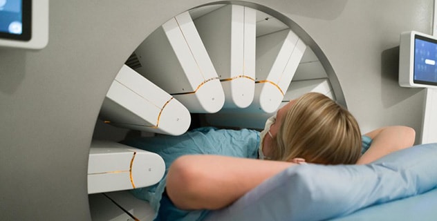
During a SPECT scan, your healthcare team positions you on a table. The camera system surrounds you and may rotate around you as it takes pictures of internal organs and other structures highlighted by the radioactive tracer in your body.
SPECT scans involve two steps:
- Receiving the tracer.
- Undergoing the SPECT scan.
Receiving the tracer. A needle is placed in a vein in your hand or arm. There may be some mild pain with the needle placement. The tracer is in a liquid that enters your blood through an IV tube attached to the needle. Your arm may feel cold when the tracer enters.
Less often, a tracer may be swallowed, inhaled or directly injected into the organ to be studied.
You may be asked to lie quietly in a room for 30 minutes or more before your scan. This gives your body time to absorb the radioactive tracer. In some cases, you may need to wait several hours or, rarely, several days between the injection and your SPECT scan.
Undergoing the SPECT scan. The SPECT machine has a circular system with several cameras that detect the radiation from the tracer. The camera system surrounds you and may rotate around you as you lie on a table.
You may be asked to change positions. During a scan you need to lie very still for certain periods of time. You may get breaks between these periods.
Children or people who are anxious about the procedure may be given medicine to help them relax. The healthcare team may include a specialist to help with staying still or coping with anxiety.
The scanning may take 20 minutes to several hours, depending on the imaging that's needed.
After
The radioactive material naturally breaks down. Most of the radioactive tracer leaves your body through urine and stool within a few hours to days after your SPECT scan. You may be told to drink plenty of water after your SPECT scan to flush the tracer from your body.
Results
SPECT scan results
SPECT scan results
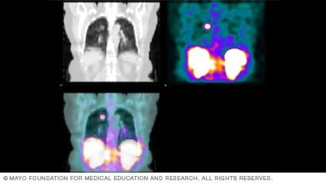
SPECT and CT scans can be combined to offer detailed imaging. The images here show a SPECT image of the liver and spleen (left), a CT image of the same view (center), and a fused SPECT and CT image (right). The different colors show different amounts of uptake of the radioactive tracer used in a SPECT scan.
A radiologist or healthcare specialist with advanced training in nuclear medicine studies the results of your SPECT scan and sends them to your healthcare team.
Ask your healthcare team how long you are likely to wait for your results.
Sept. 02, 2026