Sept. 22, 2026
Gene therapy has transformed the treatment landscape for inherited retinal diseases (IRD), offering the potential to restore or preserve vision by delivering functional genes directly to affected cells. Despite these advances, significant challenges remain. A growing body of research from Mayo Clinic investigators is exploring a novel fibrin hydrogel platform that could address some of the most persistent barriers in retinal gene and cell therapy.
Building on previous work, Mayo Clinic investigators completed a systematic review and meta-analysis evaluating the impact of retinal viral gene therapies and the prevalence of serious adverse events. The results were published in Clinical & Experimental Ophthalmology in November 2025. "We were able to conclude that there was a need for optimized delivery methods, refining dosing protocols, and improving therapeutic safety and consistency," says Brittni A. Scruggs, M.D., Ph.D., a co-author of the study and vitreoretinal surgeon at Mayo Clinic in Rochester, Minnesota.
The current standard of care for retinal gene therapy often requires subretinal injection. This procedure places gene therapy beneath the retina by creating a localized retinal detachment, often referred to as a subretinal bleb.
"While effective in targeting retinal cells, this approach treats only a limited area and carries procedural risks, particularly in patients with fragile or degenerating retinas," Dr. Scruggs says. Alternative intravitreal injections avoid retinal detachment but can dilute the therapeutic payload, reducing efficacy. Increasing the dose to overcome dilution may elevate the risk of inflammation.
Overview of FE-AAV gene therapy
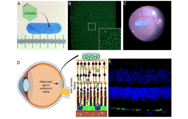
Overview of FE-AAV gene therapy
(A) Replication defective adeno-associated virus (AAV) particles (green) containing a reporter gene (GFP) are packaged in a fibrin hydrogel (blue) to create fibrin encapsulated (FE)-AAV. (B) Under the microscope, viral particles (green) can be seen in the FE-AAV gel when stained for AAV. (C) The FE-AAV is surgically implanted and "glued" to the inner surface of retina. Panel D depicts where the FE-AAV is implanted and how AAV particles traverse the retina to cause expression of the transgene in their target cells. (E) One month following FE-AAV implantation surgery, RPE is expressing green fluorescent protein.
Aiming to mitigate the limitations of current treatments, Mayo Clinic researchers developed a fibrin hydrogel therapeutic delivery. Rather than delivering gene therapy beneath the retina, investigators incorporated viral gene therapy into a thin fibrin implant that can be placed directly on the retinal surface. In a study published in Science Advances in September 2025, they introduced a fibrin hydrogel implant designed to improve gene therapy delivery within the eye. The implant is a small, biodegradable scaffold composed of fibrin, a naturally occurring protein involved in blood clotting. Because fibrin is routinely produced and degraded by the body, it represents a biologically compatible platform for therapeutic delivery.
"Through our collaborative efforts, we were able to develop a method to slow the polymerization of fibrin sufficiently to allow us to produce three-dimensionally shaped hydrogels with a high concentration of fibrin," says Alan D. Marmorstein, Ph.D., a study co-author and ophthalmology researcher at Mayo Clinic in Rochester, Minnesota. "The high concentration of fibrin is critical because it provides the stiffness that we need to manipulate it surgically combined with enough pliability to avoid damage to adjacent tissue."
One of the most notable findings reported in the study was the extent of retinal transduction achieved with the implant. As the fibrin hydrogel degrades, it releases the therapeutic agent in close proximity to target tissues while maintaining a concentrated local dose.
"Traditional subretinal approaches typically affect only the area surrounding the bleb, often representing a small fraction of the retina," Dr. Scruggs continues. "In contrast, the fibrin hydrogel system enables gene delivery across a substantially larger retinal area, extending into the peripheral retina — exceeding what is commonly achieved through localized subretinal administration."
Using the first large animal geographic atrophy model, Mayo Clinic investigators have also used the hydrogel to deliver retinal pigment epithelial cells to areas where native tissue had been intentionally removed to mimic advanced macular degeneration. The results were published in the January 2026 issue of Frontiers in Cell and Developmental Biology.
"We were interested in transplanting retinal pigment epithelial cells as a sheet," says Raymond Iezzi, Jr., M.D., a co-author of the study and a vitreoretinal surgeon at Mayo Clinic in Rochester, Minnesota. "Because a single layer of cells is not something that we can handle mechanically during surgery, we needed a scaffold to physically manipulate these cells and place them under the retina."
"We found that the treatment protected photoreceptors, improved retinal health and enhanced local blood flow, while the hydrogel safely degraded without causing significant inflammation," Dr. Marmorstein says.
Early studies suggest the platform may also have applications in other tissues and organ systems, highlighting the versatility of the fibrin hydrogel platform. Looking ahead, the Mayo Clinic research team is preparing for larger therapeutic studies and plans to advance this technology toward clinical trials.
"Fibrin hydrogel-based delivery could represent an important step forward in overcoming some of the most significant challenges facing retinal gene and cell therapy today," Dr. Iezzi says.
"Beyond these specific use cases, we're exploring the potential for our work to be adapted for nonviral gene delivery, additional cell-based therapies and other biologic treatments," Dr. Scruggs says. "Our next challenge is to investigate how these advances might translate to nonophthalmic applications."
For more information
Berger A, et al. Retinal viral gene therapy: Impact of route of administration on serious adverse events — A systematic review. Clinical & Experimental Ophthalmology. 2025;53:967.
Scruggs BA, et al. Retinal gene therapy using epiretinal AAV-containing fibrin hydrogel implants. Science Advances. 2025;11:eadv7922.
Marmorstein AD, et al. Degradable fibrin hydrogels for transplantation of iPSC-derived retinal pigment epithelial cell monolayers. Frontiers in Cell and Developmental Biology. 2026;13:1739620.
Refer a patient to Mayo Clinic.