April 24, 2026
Mayo Clinic's specialized moyamoya disease team has deep expertise in managing this challenging condition. Although rare, moyamoya disease can have devastating consequences, triggering strokes and seizures in people under age 50.
"Moyamoya is a subtle disease. At Mayo Clinic, we are at the forefront of currently available diagnostic and treatment capabilities," says Rabih G. Tawk, M.D., a neurosurgeon at Mayo Clinic in Jacksonville, Florida.
Moyamoya disease is a progressive condition characterized by blockage in the distal internal carotid artery and adjacent blood vessels. The name moyamoya — which means "puff of smoke" in Japanese — describes the appearance of the tangle of tiny blood vessels formed to compensate for the blockage. The disease generally occurs in fewer than 1 in 100,000 people. Mayo Clinic specialists evaluate and treat more than 170 patients each year with moyamoya disease.
Early diagnosis and treatment are essential to prevent debilitating strokes and seizures. But diagnosis is challenging, as moyamoya disease is often insidious and has many mimics, including migraine and multiple sclerosis.
The moyamoya disease clinic at Mayo Clinic includes specialists in neurology, neurosurgery, neuroradiology and neuropsychology. "Patients see not just a surgeon or a neurologist but an entire team that understands this disease," Dr. Tawk says. "That's what's different about Mayo Clinic." Expert management of moyamoya disease is available enterprise-wide.
State-of-the-art imaging
Dr. Tawk recommends that physicians consider referring a patient to moyamoya disease specialists if the patient has had recurrent strokes with cerebrovascular occlusive disease primarily centered at the large intracranial vessels as seen on noninvasive imaging, including CT angiography and MR angiography. Other subtle findings include unilateral vasculopathy in patients with headache and cognitive decline.
Conventional angiography best demonstrates the extent of occlusive disease and the characteristic puff of smoke appearance of small collateral blood vessels and can establish the diagnosis. But optimal management requires further evaluation by specialists experienced in moyamoya disease.
"We have seen several patients who had imaging done elsewhere and were managed for prolonged periods, but moyamoya was missed," Dr. Tawk says.
Mayo Clinic uses several state-of-the-art diagnostic technologies, including 7T MRI and photon-counting detector CT. Moyamoya diagnostic testing includes:
 Imaging that guides treatment plans
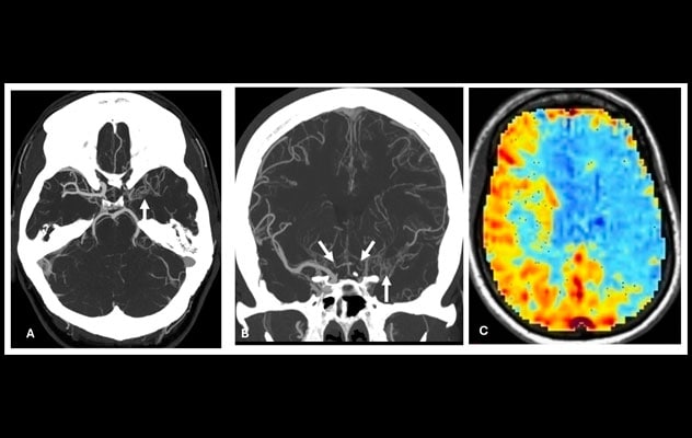
Axial (A) and coronal (B) CT angiography images of the head show moyamoya disease changes involving the left middle cerebral artery and bilateral anterior cerebral arteries (arrows). A blood oxygenation level-dependent (BOLD) functional MRI cerebrovascular reactivity color map (C) shows decreased cerebrovascular reserve within the left middle cerebral artery and bilateral anterior cerebral artery territories.
- MRI to identify infarcts and white matter changes.
- Vessel wall imaging to evaluate vessel wall thickening and enhancement.
- Perfusion MRI to measure blood flow.
- Blood oxygenation level-dependent (BOLD) functional MRI to assess cerebrovascular reserve — sometimes called a brain stress test.
"Instead of relying solely on structural changes, we can measure cerebral blood flow, vascular reserve and functional impact. These technologies reduce diagnostic uncertainty," says Richard D. Beegle, M.D., a neuroradiologist at Mayo Clinic's campus in Florida. "For patients with moyamoya vasculopathy, being treated at a center with dedicated neurovascular imaging expertise ensures that complex imaging studies are interpreted accurately and in context. Because the disease is rare, experience matters — both in acquiring the studies and in understanding what they truly mean for each individual patient."
Additional diagnostic evaluations might include vision checks and neuropsychological testing, to assess memory and cognition. "All of these test results are important to determining personalized treatment," Dr. Tawk says. "Delivering the best care requires the best possible information."
Optimizing outcomes
The first line of treatment for moyamoya disease is medication to control symptoms and reduce the risk of stroke. Surgery is considered the gold standard treatment and is recommended when patients have a history of strokes, symptoms of cerebrovascular insufficiency or impaired blood flow to the brain.
"Patients see not just a surgeon or a neurologist but an entire team that understands this disease."
Mayo Clinic neurosurgeons typically use combined direct and indirect bypass techniques for revascularization, to optimize outcomes while minimizing complications. Neuroanesthesiologists and neurocritical care specialists provide intraoperative and postoperative management to further support optimal outcomes.
Patients are followed longitudinally in the moyamoya clinic with repeat imaging to assess disease progression and bypass maturation, to learn if the risk of stroke has eased. "We have many patients we have followed for more than 10 years. They had their surgery and they're doing great," Dr. Tawk says.
Optimal outcomes require collaborative expertise. "A condition as challenging as moyamoya disease requires a multidisciplinary approach, like a chain where every ring is needed," Dr. Tawk says. "Even the best surgeon will not have good outcomes without high-quality imaging, an anesthesiologist who watches these patients like a hawk, and a critical care expert to manage these patients after surgery.
"We don't want this subtle disease to disable patients," Dr. Tawk says. "Our expertise and technology are aimed at giving people a good quality of life."
For more information
Refer a patient to Mayo Clinic.