Jan. 20, 2018
Computer-Aided Nodule Assessment and Risk Yield (CANARY) is a novel image analysis software application. It was developed to noninvasively predict the histology and risk stratify pulmonary nodules of the lung adenocarcinoma spectrum, which comprises almost all indolent lung cancers.
CT- and CANARY-analyzed pulmonary nodule
CT- and CANARY-analyzed pulmonary nodule
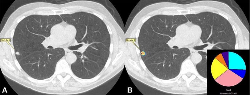
A. An indeterminate pulmonary nodule in the right upper lobe on axial noncontrast CT of the chest. B. The same nodule as coded by CANARY-identified exemplar with a representative glyph showing the proportional presence of each exemplar in inset. This nodule is stratified into the intermediate risk category.
CANARY analysis is based on the radiological density and texture of the analyzed nodule. CANARY detects and quantifies the presence of nine different recurring adenocarcinoma tissue types (exemplars) and displays a glyph portraying the proportion of nodule volume occupied by each exemplar.
Based on the proportional distribution of these exemplars, CANARY allows the classification of indeterminate and screen-detected nodules into histopathologic categories:
- Adenocarcinoma in situ
- Minimally invasive adenocarcinoma
- Invasive adenocarcinoma
Independent clustering of the CANARY glyphs stratifies these lesions into three distinct prognostic categories: good, intermediate and poor. Good nodules demonstrate an essentially 100 percent post-resection disease-free survival in early-stage disease. CANARY-based risk stratification has been externally validated in multiple data sets, including the National Lung Screening Trial.
Clinical application of CANARY
At Mayo Clinic, CANARY is routinely used in Pulmonary and Critical Care Medicine's Lung Nodule, Mass and Adenopathy Clinic and during multidisciplinary lung cancer tumor board conferences.
To individualize patient management, CANARY data are specifically applied in challenging cases of multifocal adenocarcinoma of the lung, and in solitary nodules in high-risk surgical candidates. Glyph stability and change in composition is incorporated into joint decision-making with the patient. While further investigation regarding the longitudinal changes of these lesions is needed, researchers anticipate CANARY will become a very valuable tool in these complex cases.
Ongoing research
CANARY recently has shown promise in its ability to noninvasively predict epithelial growth factor receptor (EGFR) status of adenocarcinoma of the lung. This application could allow better selection of subjects for EGFR-targeted therapy without necessitating biopsy.
Differentiation of benign and malignant pulmonary nodules
Funded by the Department of Defense, a multidisciplinary team of researchers from Mayo Clinic and Vanderbilt University used the National Lung Screening Trial database to develop a radiomic model to differentiate benign from malignant screen-detected and incidentally detected pulmonary nodules ranging in size from 7 to 30 mm.
Multivariate analysis identified several variables — including nodule location, size, shape, density, texture and surface characteristics — that differentiated benign from malignant pulmonary nodules, with an area under the curve of 0.94. This model has been internally validated and external validation is ongoing.