March 24, 2026
Neurosurgeons at Mayo Clinic in Phoenix/Scottsdale, Arizona, generally avoid the use of felt padding when treating trigeminal neuralgia. Over time, those polytetrafluoroethylene spacers — intended to relieve a blood vessel's contact on the trigeminal nerve root — can themselves cause problems, leading to recurrent symptoms.
 Compression after decompression
Intraoperative image shows discoloration of a granuloma and inflammation that can develop after conventional trigeminal nerve decompression using a felt implant. The inflammation is directly against the nerve root in a patient with recurrent pain.
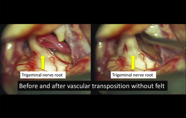 Decompression without felt
On the left, an initial intraoperative image shows a blood vessel compressing the trigeminal nerve root. On the right, post-transposition image shows the blood vessel surgically moved away from the nerve root, avoiding the use of felt.
"Patients can initially have good outcomes when felt is placed. But in unfortunate cases, the pain comes back," says Richard S. Zimmerman, M.D., a neurosurgeon at Mayo Clinic's campus in Arizona. "Our research has shown that chronic inflammation can develop in the implant over five or 10 years. A procedure meant to decompress the nerve can end up causing both inflammation and ongoing compression."
Mayo Clinic's approach involves surgically moving the offending blood vessel off the nerve — leaving it untouched — and then using fibrin glue or other options to anchor the blood vessel to a new site.
"As the fibrin glue naturally breaks down, it's replaced by arachnoid scar tissue. It's the patient's own body tissue that keeps the blood vessel stuck to its new location," Dr. Zimmerman says. "This procedure is more nuanced and innovative compared to the typical microvascular decompression." Mayo Clinic's experience with the procedure was described in Operative Neurosurgery.
Mayo Clinic offers several options for the treatment of trigeminal neuralgia. Specialists in neurology, radiation oncology, interventional neuroradiology, pain management and rehabilitation, and neurosurgery work together to provide precise diagnosis of the cause of facial pain, and personalized treatment.
"At Mayo Clinic, you have more brains thinking about what's wrong — and more options," Dr. Zimmerman says.
Key steps in the diagnosis of facial pain
Determining the cause of facial pain can be challenging. In addition to trigeminal neuralgia, the possibilities include malignancy and dental issues.
Dr. Zimmerman recommends asking patients if they experienced facial numbness before facial pain. "Numbness isn't typical for trigeminal neuralgia," he says. "That symptom should raise suspicions of malignancy, especially if the patient has a prior history of skin cancer of the face or scalp."
Dental issues and trigeminal neuralgia can be difficult to differentiate. Pain associated with dental issues typically is continuous and described as throbbing or aching. "But the pain most commonly seen in trigeminal neuralgia has acute attacks that come and go. It's frequently described as stabbing or like an electric shock," Dr. Zimmerman says. "It might be triggered by wind in the face, showering or some other facial stimulation that is seemingly innocuous, such as applying makeup or shaving."
Patients with trigeminal neuralgia often first consult a dentist or endodontist because the pain seems to originate in the mouth or teeth. Dr. Zimmerman notes that Mayo Clinic specialists have seen patients with trigeminal neuralgia who had multiple unnecessary procedures before receiving a correct diagnosis. "If a diagnosis for the cause of orofacial pain is uncertain or an initial treatment doesn't work, providers should think about getting a second opinion, usually from a neurologist," Dr. Zimmerman says.
When trigeminal neuralgia is suspected, Mayo Clinic uses cutting-edge 7T MRI to assist in the diagnosis. Mayo Clinic has a custom trigeminal neuralgia MRI protocol, designed by Mayo Clinic physicists and neuroradiologists through extensive volunteer scanning and clinical validation.
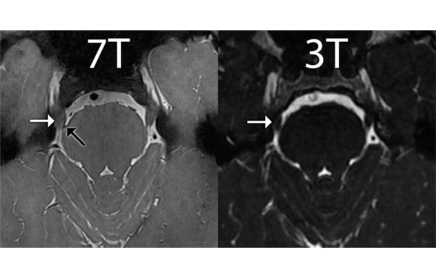 Clarity at 7T
On the left, 7T proton density MRI sequence shows the tiny superior cerebellar artery branch (black arrow) compressing the trigeminal nerve (white arrow). On the right, that tiny vessel isn't resolved at 3T MRI.
"The vascular structures pushing on the trigeminal nerve can be very small. Our 7T imaging protocol doubles the spatial resolution and decreases image noise compared with 3T MRI, leading to improved visualization of the trigeminal nerve and adjacent vascular structures," says Justin Cramer, M.D., a neuroradiologist at Mayo Clinic's campus in Arizona. "Improved visualization of this region increases the likelihood of a correct diagnosis and helps guide treatment decisions."
Imaging with 18F-fluorodeoxyglucose (18F-FDG) positron emission tomography (PET) provides metabolic insight that can indicate active inflammation. 18F-FDG PET imaging is especially pertinent for patients with felt implants from prior surgery.
"We have identified increased pathological metabolism using PET in the areas where our MRI identifies an implant," Dr. Zimmerman says. Mayo Clinic researchers described the correlation between 18F-FDG PET imaging and pathology in Clinical Nuclear Medicine.
Nonsurgical treatment
Medications can manage the symptoms of trigeminal neuralgia. However, patients often experience significant side effects, and the medication's efficacy can decrease over time. "This is clearly problematic for any patient that has to commit to lifelong medication, probably at an increasing dose, that brings with it confusion, fatigue and impaired executive function," Dr. Zimmerman says.
Mayo Clinic's interventional neuroradiologists provide treatment for patients who wish to avoid medication and aren't surgical candidates. Extracranial nerve blocks are one option. Other treatments — such as intracranial glycerol injections and noninvasive stereotactic radiosurgery — selectively damage the trigeminal nerve to block pain. Specialists in pain rehabilitation also can provide physical and cognitive therapies to help improve patients' quality of life.
Mayo Clinic's commitment to managing trigeminal neuralgia entails investments in technology and surgical expertise. Dr. Zimmerman serves on the medical advisory board of the Facial Pain Association, a patient advocacy group.
"Our multidisciplinary care focuses on what is best for the patient. That's the core of the Mayo Clinic Model of Care," Dr. Zimmerman says.
For more information
Pines AR, et al. Microvascular transposition without Teflon: A single institution's 17-year experience treating trigeminal neuralgia. Operative Neurosurgery. 2021;20:397.
Herber S, et al. Teflon granuloma with active inflammation: A cause of recurrent trigeminal neuralgia after microvascular decompression — Imaging and pathological correlation. Clinical Nuclear Medicine. 2024;49:1105.
Facial Pain Association.
Refer a patient to Mayo Clinic.