Sept. 15, 2026
Interest has grown in the pulvinar as a potential neurostimulation target for drug-resistant epilepsy. Mayo Clinic researchers have mapped the influence of pulvinar subregions on select cortical networks in humans. The detailed maps have potential to guide the further development of pulvinar subregions as distinct neuromodulation targets.
To causally map the pulvinar's influence on the cortex, the researchers measured brain stimulation-evoked potentials in 30 patients with drug-resistant epilepsy. The patients were implanted with stereoelectroencephalography with pulvinar electrode coverage during clinically indicated epilepsy monitoring at Mayo Clinic in Rochester, Minnesota, or Barnes-Jewish Hospital in St. Louis, Missouri. The study results were published in the Journal of Neuroscience.
Key findings:
- Stimulation of the lateral pulvinar influenced striate and extrastriate regions.
- Ventral-medial pulvinar stimulation preferentially influenced lateral temporal cortices.
- Dorsomedial pulvinar stimulation influenced the parietal cortex.
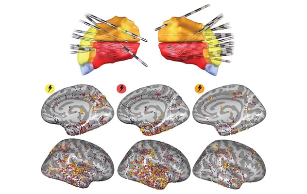 Stimulating pulvinar subregions
Illustration shows which parts of the brain respond to stimulation of various regions of the pulvinar. On the left, stimulating the outer (lateral) part of the pulvinar primarily activates areas involved in vision (yellow). In the middle, stimulating the lower middle (ventral-medial) pulvinar activates brain regions involved in memory, language and object recognition (red). On the right, stimulating the upper middle (dorsal-medial) pulvinar activates regions involved in attention, spatial awareness and planning (orange).
These effects were captured by a compact set of large-scale cortical gradients, including outputs along a dorsal-ventral, anterior-posterior and medial-lateral pulvinar axis. The resulting causal map summarizes pulvinar subregion influence on the cortex with gradients that depict its large-scale organization.
"We were surprised by how large, detailed and complex this deep brain structure is, and by its potential role in guiding epilepsy treatments," says Dora Hermes Miller, Ph.D., a biomedical engineer at Mayo Clinic's campus in Minnesota and the study's senior author.
The study demonstrated that pulvinar regions separated by as little as 3 millimeters connected to completely different brain networks. "That level of detail means that if neurologists and neurosurgeons want to suppress the seizures coming from those areas, they have to place electrodes in the precise right spot," Dr. Hermes says. "These findings provide a guide towards that spot."
The pulvinar maps have immediate relevance for clinical care. "We are already using these maps to help individualize pulvinar deep brain stimulation targeting in patients with drug-resistant epilepsy, while continuing to study how these maps relate to long-term outcomes," says Nick M. Gregg, M.D., a neurologist at Mayo Clinic's campus in Minnesota and a study co-author.
The work also furthers Mayo Clinic's commitment to individualized care for drug-resistant epilepsy. "These findings provide data that enables exploration of tailoring neuromodulation therapies in a more personalized way, targeting each patient's specific epilepsy networks," says Gregory A. Worrell, M.D., Ph.D., a neurologist at Mayo Clinic's campus in Minnesota and a study co-author.
Research continues into which parts of the pulvinar should be stimulated, and at what frequencies, to better control seizures while minimizing side effects. "Our aim is to make therapy more precise, more consistent and ultimately more effective," Dr. Gregg says.
For more information
Bilderbeek JA, et al. Pulvinar subregions influence select cortical pathways in humans. Journal of Neuroscience. 2026;46: e1604252026.
Refer a patient to Mayo Clinic.