Overview
Pituitary gland magnetic resonance imaging (MRI) uses a strong magnetic field and radio waves to make detailed pictures of the pituitary gland and nearby areas. This small, pea-sized gland is located behind the nose at the base of the brain. It makes hormones that help control many important body functions.
This imaging test may be used to look for a tumor, called a pituitary adenoma, or another change in or near the pituitary gland. A pituitary MRI can help show the size and shape of the gland. It can help show whether a growth is pressing on nearby areas. It also can help show whether an existing finding has changed over time.
A pituitary gland MRI is different from a standard brain MRI. It uses images focused on the pituitary area, often with thinner slices. Contrast material may be used to help see things more clearly.
Why it's done
Pituitary tumor
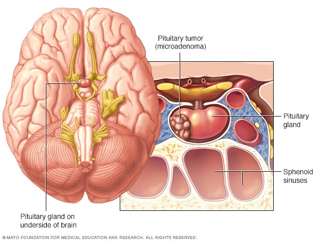
Pituitary tumor
Pituitary tumors are tumors that form in the pituitary gland at the base of the brain. These tumors can cause changes in hormone levels. This illustration shows a smaller tumor, called a microadenoma.
A pituitary gland MRI focuses on the pituitary gland and nearby areas. These include the sella, the optic chiasm, the pituitary stalk and the cavernous sinuses. The sella, also called the pituitary fossa, is the bony space that holds the pituitary gland. The optic chiasm is where the optic nerves cross. The pituitary stalk, also called the infundibulum, connects the pituitary gland to the brain. The cavernous sinuses are spaces near the pituitary gland that contain important nerves and blood vessels.
A pituitary MRI may be used to:
- Look for a pituitary tumor or another change in tissue in or near the pituitary gland. Most pituitary tumors are not cancer. But they can cause symptoms if they make too much hormone or press on nearby areas.
Pituitary tumors may be described as microadenomas or macroadenomas based on their size. A microadenoma is smaller than 10 millimeters. A macroadenoma is 10 millimeters or larger. For comparison, 10 millimeters is about 1/2 inch. Small tumors may be harder to see than larger tumors. Contrast material can help show some small tumors more clearly.
- Evaluate symptoms. Symptoms that may be related to a pituitary growth or hormone changes include:
- Headaches.
- Vision changes, including double vision or loss of side vision.
- Menstrual changes.
- Breast milk production not related to breastfeeding.
- Sexual function changes, such as lower interest in sex, erectile dysfunction or fertility issues.
- Find out why blood test results suggest the pituitary gland may be making too much or too little hormone. These results may involve prolactin, growth hormone, cortisol, thyroid hormones or sex hormones. Low testosterone may lead to pituitary testing if the pattern of blood test results suggests that the pituitary gland may not be sending the expected signals to the testicles.
- Examine a pituitary finding found by chance during a brain MRI or CT scan done for another reason. This is called a pituitary incidentaloma. These findings may include an enlarged pituitary gland, thickening or shifting of the pituitary stalk, a cyst, or an empty sella. An empty sella means the bony space that holds the pituitary gland looks partly or mostly empty because the gland appears flattened.
- Monitor a known pituitary finding over time.
- Check how treatment is working. After medicine, surgery or radiation therapy, a pituitary MRI may show how the pituitary gland and nearby areas have changed. It also may help the healthcare team look for signs that a growth has come back.
Brain MRI versus pituitary gland MRI
A standard brain MRI looks broadly at the brain. A pituitary gland MRI focuses on the pituitary gland and the small structures nearby. It often uses thinner image slices through the pituitary area. These focused images can show small pituitary findings that may be hard to see on a routine brain MRI.
A pituitary gland MRI often includes images taken from more than one angle. The MRI also may use contrast material, which can help show a small lesion more clearly.
A dedicated pituitary MRI may not be needed in every situation. For example, if a previous scan already gives enough information, the healthcare team may not need another MRI right away.
Risks
MRI does not use X-rays or ionizing radiation. But the MRI machine uses a strong magnet, so safety screening is important before the test. Some metal objects, implanted devices and medical equipment can be affected by the magnet. The healthcare team checks for these risks before the scan.
Some implanted devices are safe for MRI under certain conditions. Others may need special planning, device checks or a different imaging test. Tell the healthcare team if you have an implanted medical device or metal in your body.
Some people feel anxious or uncomfortable in the MRI scanner. The scanner also makes loud knocking or tapping sounds during the test. Ear protection typically is provided.
Contrast material
Most pituitary gland MRI exams use contrast material. It typically is given through an IV in a hand or an arm. Contrast material can help healthcare professionals see small pituitary lesions and better understand a finding in or near the pituitary gland.
Reactions to gadolinium-based MRI contrast material are not common. When reactions happen, they often are mild. Serious reactions are rare. Tell the healthcare team if you have ever had a reaction to MRI contrast material. A past reaction to the same type of contrast material is one of the strongest risk factors for another reaction.
Some people notice mild side effects after contrast material, such as nausea, headache, dizziness, itching or a rash. Tell the healthcare professional right away if you feel short of breath, have swelling, feel lightheaded or have other symptoms during or after the injection.
Kidney function may need to be checked before MRI contrast material is used, especially for people with kidney disease. This is because some gadolinium-based contrast materials have been linked to a rare condition called nephrogenic systemic fibrosis in people with severely reduced kidney function. But this risk is very low with the contrast materials most commonly used today.
The healthcare team decides whether contrast material is needed based on the reason for the scan and the type of pituitary finding being evaluated. The team also considers kidney function, pregnancy status and any past reactions to contrast material.
How you prepare
Before a pituitary gland MRI, follow the instructions from the imaging center. You may need to complete an MRI safety form before the test. Answer all questions carefully.
Bring your implanted device card if you have one. This can help the healthcare team confirm the type of device and whether it is safe to scan in an MRI machine.
You may need to change into clothing that the imaging center gives you. Leave jewelry, watches, hairpins and other metal items at home, or remove them before the scan.
Tell the healthcare team before the scan if you are pregnant or think you might be pregnant. Also tell the team if you have kidney disease or have had a reaction to MRI contrast material in the past.
You usually do not need to stop eating or drinking before a routine MRI with contrast material. This may be different if you will receive medicine to help you relax or if anesthesia is planned. Follow what your healthcare team tells you.
If you feel anxious in small spaces, tell the healthcare team before the day of your test. The team can discuss ways to help you feel more comfortable during the MRI.
What you can expect
Pituitary gland MRI is usually done in an MRI suite at a hospital or imaging center. The test may be done with or without contrast material.
Before the test
The imaging center may give you clothing to change into before the test. The healthcare team reviews your MRI safety form. The team asks you about metal, implanted devices, pregnancy, kidney disease, past contrast reactions and anxiety in enclosed spaces.
If contrast material is used, a member of the healthcare team places an IV in your hand or arm. The contrast material is given through the IV during the test.
During the test
MRI scanner room and viewing room
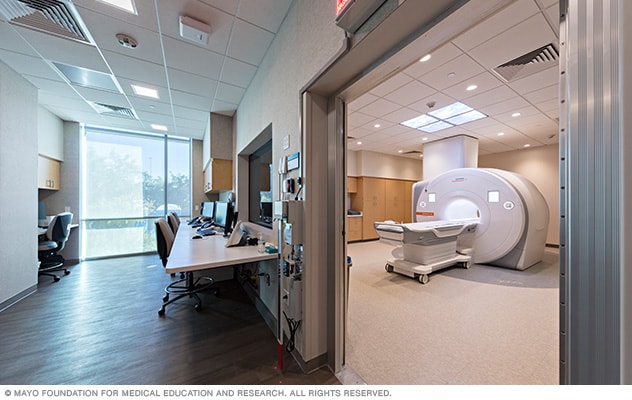
MRI scanner room and viewing room
The MRI scanner is in a separate room from the technologists who operate the machine and capture the images of your pituitary gland. You can communicate between the rooms through a speaker.
You lie on a padded table that slides into the MRI scanner. Your head may be supported with cushions or a frame to help you stay still. Staying still helps the MRI create clear pictures.
The scanner makes loud tapping, thumping or knocking sounds while it takes pictures. You usually receive ear protection. You may be able to talk with the technologist through an intercom during the test.
A pituitary gland MRI often includes images taken from several directions. These may include coronal, sagittal and axial views. Coronal views show the gland from front to back. Sagittal views show the gland from the side. Axial views show the gland from top to bottom.
Some pituitary MRI exams use dynamic contrast-enhanced MRI. This means the scanner takes repeated images as contrast material moves through the pituitary gland. Dynamic imaging can help healthcare professionals evaluate some small pituitary lesions.
If contrast material is used, you may feel a cool sensation when it is injected. Some people notice a brief taste in the mouth or mild nausea. Tell the technologist right away if you feel short of breath, itchy, lightheaded or unwell.
The amount of time the scan takes can vary. Many pituitary gland MRI exams take about 30 to 60 minutes. The scan may take longer if more images are needed or if sedation is used.
After the test
Most people can return to their regular activities after the MRI. If you received anesthesia or medicine to help you relax, you may need time to recover. You also may need someone to drive you home.
If you had contrast material, the IV is removed after the test. Tell the healthcare team if you notice symptoms after leaving the imaging center. These may include a rash, itching, swelling or trouble breathing.
Results
A radiologist reviews the pituitary gland MRI images and sends a report to the healthcare professional who ordered the test. Your healthcare team explains what the results mean and whether you need more testing, monitoring or treatment.
A pituitary gland MRI report may describe:
Pituitary adenomas and other lesions
A pituitary adenoma may appear as a small area that looks different from the rest of the pituitary gland, especially after contrast material is given. A microadenoma is smaller than 10 millimeters and may be hard to see. A macroadenoma is 10 millimeters or larger. A larger adenoma may change the shape of the sella, press upward toward the optic chiasm or extend toward the cavernous sinuses.
The report may describe whether contrast material makes an area brighter in an even or uneven pattern. When the pattern is uneven, it is called heterogenous enhancement. This finding does not point to one diagnosis by itself.
Some pituitary findings can be seen without contrast material, especially larger lesions or changes in the shape of the gland. Contrast material can help show smaller lesions and better define a finding in or near the pituitary gland.
A cystic lesion is a fluid-filled finding. Cystic lesions in or near the pituitary gland may include Rathke cleft cysts or cystic pituitary adenomas. Some cystic findings may not need treatment and instead may be monitored with follow-up MRI.
Other pituitary gland MRI findings
The report may note an enlarged pituitary gland. This can have many causes. An enlarged gland may be related to a pituitary adenoma, pituitary hyperplasia, inflammation, pregnancy-related change or another condition. Pituitary hyperplasia means the gland is enlarged because pituitary cells have increased in number. This can happen during pregnancy or when hormone signals cause the pituitary gland to work harder.
The report also may mention the posterior pituitary bright spot. This is a bright area that is often seen in the back part of the pituitary gland on certain MRI images. The bright spot may become less visible with age. It also may not be seen in people with arginine vasopressin (AVP) deficiency, also called central diabetes insipidus, or with some posterior pituitary lesions.
The report may describe the pituitary stalk, which connects the pituitary gland to the brain. Stalk deviation means the stalk is shifted to one side. This can happen when a lesion changes the shape of the gland. Stalk thickening means the stalk looks wider than expected and may need more evaluation.
An empty sella means the sella looks partly or mostly filled with fluid. This can make the pituitary gland look flattened along the bottom of the sella. An empty sella is often found by chance and may not cause symptoms. This finding does not point to one specific condition by itself. It may be related to past pituitary changes or conditions associated with increased intracranial pressure. The meaning depends on symptoms, hormone test results and the rest of the MRI findings.
Findings that may need follow-up
A pituitary MRI finding may need closer evaluation if it is growing, affecting hormone levels or pressing on nearby structures such as the optic chiasm or cavernous sinuses. But not every pituitary finding needs treatment right away. Some findings can be watched over time with follow-up MRI exams, blood tests, vision testing or visits with specialists.
Prompt evaluation may be needed if MRI shows bleeding, tissue damage from loss of blood flow, or pressure on nearby structures. Pituitary hemorrhage means bleeding in the pituitary gland or in a pituitary tumor. When sudden bleeding or loss of blood flow occurs in a pituitary adenoma, it can cause a condition called pituitary apoplexy. Pituitary apoplexy is a medical emergency that needs urgent medical evaluation. Symptoms may include sudden severe headache, nausea, vomiting, vision changes, double vision, new hormone issues or decreased alertness.
Clinical trials
Explore Mayo Clinic studies of tests and procedures to help prevent, detect, treat or manage conditions.
Aug. 20, 2026