Retinal detachment Symptoms and causes
Overview
Retinal detachment happens when the thin layer of tissue at the back of the eye pulls away from its regular position. This layer of tissue is called the retina. Retinal detachment is an emergency.
Retinal detachment separates the retinal cells from the layer of blood vessels that provides oxygen and nourishment to the eye. The longer retinal detachment goes without treatment, the greater the risk of permanent vision loss in the affected eye.
Symptoms of retinal detachment can include the following: reduced vision, the sudden appearance of dark floating shapes and flashes of light in your vision, and loss of side vision. Contacting an eye doctor, called an ophthalmologist, right away can help save your vision.
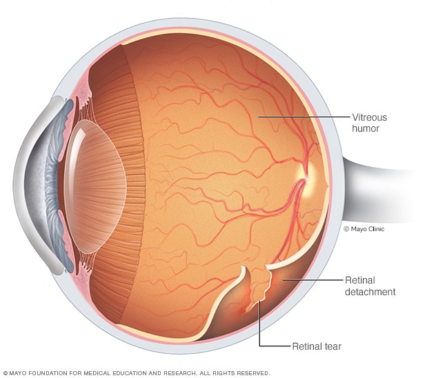
Retinal detachment
Retinal detachment describes an emergency in which a critical layer of eye tissue called the retina pulls away from the layer of blood vessels that provide it with oxygen and nutrients. The retina is at the back of the eye. Retinal detachment often comes with flashes and floaters in your vision.
Symptoms
Retinal detachment is painless. Often, symptoms are present before a retinal detachment happens or before it has gotten worse. You may notice symptoms such as:
- The sudden appearance of tiny specks or squiggly lines that seem to drift through your field of vision. These are called floaters.
- Flashes of light in one or both eyes. These are called photopsias.
- Blurred vision.
- Side vision, also called peripheral vision, that becomes worse.
- A curtainlike shadow over your field of vision.
When to see a doctor
See a healthcare professional right away if you have any symptoms of retinal detachment. This condition is an emergency that can cause lasting vision loss.
Causes
There are three main types of retinal detachment, and their causes vary:
-
Rhegmatogenous (reg-mu-TOJ-uh-nus). This type of retinal detachment is the most common. A rhegmatogenous detachment is caused by a hole or tear in the retina that lets fluid pass through and collect underneath the retina. This fluid builds up and causes the retina to pull away from underlying tissues. The areas where the retina detaches lose their blood supply and stop working. This causes you to lose vision.
The most common cause of rhegmatogenous detachment is aging. As you age, gel-like material that fills the inside of your eye, called vitreous (VIT-ree-us), may change in texture and shrink or become more liquid. Usually, the vitreous separates from the surface of the retina without any complications. This is a common condition called a posterior vitreous detachment (PVD).
As the vitreous separates or peels off the retina, it may tug on the retina with enough force to create a tear. Most of the time it doesn't. But if a PVD causes a tear and the tear isn't treated, the liquid vitreous can pass through the tear into the space behind the retina. This causes the retina to detach.
- Tractional. This type of detachment can happen when scar tissue grows on the retina's surface. The scar tissue causes the retina to pull away from the back of the eye. Tractional detachment usually is seen in people who have poorly controlled diabetes.
- Exudative. In this type of detachment, fluid builds up beneath the retina, but there are no holes or tears in the retina. Exudative detachment can be caused by age-related macular degeneration, infection, tumors or inflammatory conditions.
Risk factors
The following factors raise your risk of retinal detachment:
- Aging — retinal detachment is more common in people ages 40 to 70.
- Past retinal detachment in one eye.
- Family history of retinal detachment.
- Extreme nearsightedness, also called myopia.
- Past eye surgery, such as cataract removal.
- Past severe eye injury.
- History of other eye disease or condition, including retinoschisis, uveitis or thinning of the peripheral retina called lattice degeneration.
From Mayo Clinic to your inbox
Sign up for free and stay up to date on research advancements, health tips, current health topics, and expertise on managing health. Click here for an email preview.
CON-20156916
Mayo Clinic does not endorse companies or products. Advertising revenue supports our not-for-profit mission.
Advertising & Sponsorship
Mayo Clinic Press
Check out these best-sellers and special offers on books and newsletters from Mayo Clinic Press.
