Descripción general
La doble salida del ventrículo derecho es una afección cardíaca presente en el momento del nacimiento. Eso significa que es un defecto cardíaco congénito. Cuando esta afección está presente, la cavidad inferior derecha del corazón tiene dos aberturas para que la sangre salga del corazón en lugar de una sola. Parte de la sangre de la cavidad inferior derecha del corazón va al cuerpo en lugar de los pulmones.
Los bebés con doble salida del ventrículo derecho también tienen un orificio entre las cavidades inferiores del corazón. Las cavidades cardíacas inferiores se llaman ventrículos. El orificio se denomina defecto septal ventricular. El orificio hace que la sangre rica en oxígeno se mezcle con sangre poco oxigenada. Los bebés que tienen este orificio pueden no recibir suficiente oxígeno en el torrente sanguíneo. Su piel puede ser grisácea o azulada.
La doble salida del ventrículo derecho puede ocurrir junto con otras afecciones cardíacas presentes en el nacimiento, como otros orificios en el corazón y los vasos sanguíneos o cambios en las válvulas cardíacas.
Doble salida del ventrículo derecho
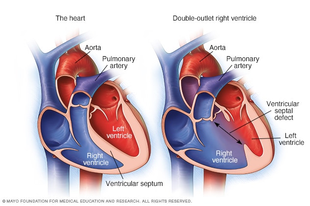
Doble salida del ventrículo derecho
En cuanto al ventrículo derecho de salida doble, la cavidad inferior derecha del corazón, denominada ventrículo derecho, tiene dos aberturas para la salida de la sangre en lugar de una. Parte de la sangre va al cuerpo en lugar de solo a los pulmones. También hay un orificio entre las dos cavidades inferiores del corazón. El orificio se denomina defecto septal ventricular. En un corazón típico, que se muestra a la izquierda, la arteria pulmonar se conecta al ventrículo derecho y la aorta se conecta al ventrículo izquierdo.
Types
There are several types of double-outlet right ventricle (DORV). The difference between these types is the location of the ventricular septal defect (VSD) and whether it is close to or far from the pulmonary artery and the aorta. The type of DORV determines which treatment may be right for your child.
Subaortic VSD
With subaortic VSD, the opening between the ventricles is close to the aorta. Because both arteries connect to the right ventricle in DORV, the left ventricle does not have its usual direct path to the aorta.
Subpulmonary VSD and Taussig-Bing type
With subpulmonary VSD, the opening between the ventricles is close to the pulmonary artery. Because of this, blood from the left ventricle tends to flow toward the pulmonary artery instead of the aorta. Taussig-Bing type is a form of DORV with this type of VSD.
Doubly committed VSD
With doubly committed VSD, the opening between the ventricles is close to both the aorta and the pulmonary artery. Because of its location, blood from the left ventricle can reach either artery.
Noncommitted VSD
With noncommitted VSD, also called remote VSD, the opening between the ventricles is farther from both the aorta and the pulmonary artery.
Fallot-type DORV
With Fallot-type (fa-LOW type) DORV, the VSD is usually close to the aorta or close to both the aorta and pulmonary artery. The pathway that carries blood from the right ventricle to the pulmonary artery may be narrowed. This narrowing lessens the amount of blood that can reach the lungs.
This is called Fallot-type DORV because it can resemble tetralogy of Fallot, which is another congenital heart defect that can lessen blood flow to the lungs.
Productos y servicios
Síntomas
Síntomas de la doble salida del ventrículo derecho:
- Labios, lengua o uñas de color grisáceo pálido o azulado. Según el color de piel, estos cambios pueden ser más difíciles o más fáciles de notar.
- Respiración rápida.
- Problemas con la alimentación, el crecimiento y el aumento de peso.
Cuándo debes consultar a un médico
Si crees que tu bebé podría tener síntomas de una afección cardíaca, programa una cita médica para un control de inmediato.
Causas
Se desconoce la causa exacta de la doble salida del ventrículo derecho. Esta afección se produce cuando el bebé o feto se está desarrollando antes del nacimiento.
Sin embargo, es posible que los cambios en los genes, algunos medicamentos o ciertas enfermedades, y los factores ambientales o del estilo de vida desempeñen un papel importante. Fumar durante el embarazo también puede aumentar el riesgo de ciertos defectos cardíacos congénitos en el feto.
En general, la arteria principal del cuerpo, llamada aorta, se conecta con la cavidad inferior izquierda del corazón y la arteria pulmonar se conecta con la cavidad inferior derecha del corazón. Pero en los bebés con doble salida del ventrículo derecho, tanto la aorta como la arteria pulmonar se conectan de manera total o parcial con la cavidad inferior derecha del corazón.
Factores de riesgo
Tener antecedentes familiares de afecciones cardíacas congénitas puede aumentar el riesgo de defectos cardíacos congénitos, como la doble salida del ventrículo derecho.
Complicaciones
Las complicaciones de la doble salida del ventrículo derecho incluyen problemas de crecimiento e insuficiencia cardíaca.
Sept. 10, 2026