Birthmarks are spots or areas of skin that look different from the skin around them. They may appear at birth, or some may develop weeks later. Types include café au lait spot, congenital nevus and hemangioma.
Birthmarks are usually harmless and don't need treatment. Some fade with time. People may seek treatment for cosmetic reasons or because the birthmark is painful, large or fast growing. Some birthmarks may be a sign of an underlying condition.
Types
There are many types of birthmarks. They can differ in color, size, shape and where they appear on the body. Some types include:
Cafe au lait spot
Cafe au lait (ka-FAY o lay) spots are flat oval or round patches on the skin. They range in color from tan to dark brown. They are permanent and very common. Cafe au lait spots can occur anywhere on the body, and the size increases as the child grows. Usually, no treatment is needed.
Children may have one or several cafe au lait spots. Having several spots may be a sign of a genetic condition, such as neurofibromatosis type 1. Or it may be a sign of some other serious condition. Talk with a healthcare professional about whether your child should be evaluated by a dermatologist, a doctor who specializes in skin conditions.
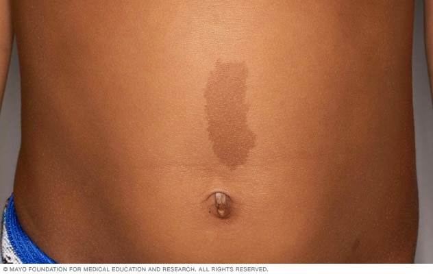
Cafe au lait spot on the abdomen.
Congenital nevus
A congenital nevus is a dark-colored mole that usually is present at birth. It often appears on the scalp or trunk of the body. It can range in size from a couple of millimeters to many centimeters in diameter. Sometimes, hair grows within the birthmark.
Children with a congenital nevus may have a higher risk of getting skin cancer later in life. The risk is greater if the birthmark is large. If your child has this type of birthmark, talk with a healthcare professional about watching for changes in the skin. Treatment may be considered based on the size and location of the birthmark, and the risk of skin cancer.
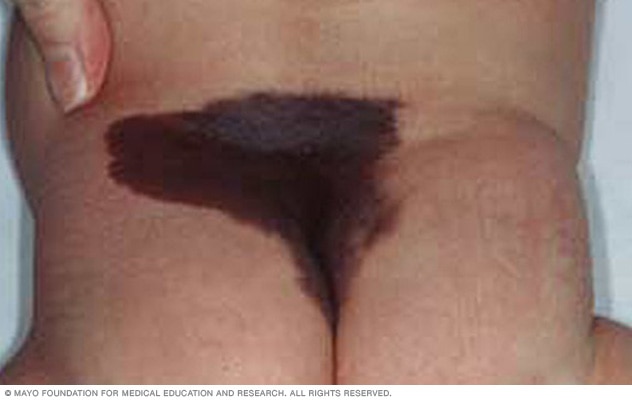
Congenital nevus on the lower back and buttocks.
Slate gray nevus
A slate gray nevus is a large flat patch. It may appear on the lower back, buttocks or shoulders. And the color ranges from deep brown to slate gray or blue-black. A slate gray nevus can be mistaken for a bruise. This type of birthmark is more common in Black, Native-American, Hispanic and Asian babies than in white babies. A slate gray nevus tends to fade during early childhood and needs no treatment.
It also is called a congenital dermal melanocytosis and was formerly called a Mongolian spot.
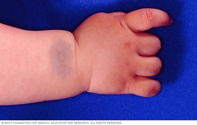
Slate gray nevus on an infant’s wrist.
Port-wine stain
A port-wine stain is a permanent birthmark present at birth. It may look pink, or light brown to deep maroon. The birthmark grows larger as the child grows. Most often, a port-wine stain appears on the face, but it can affect other areas. Over time, the affected skin may become thicker and darker.
Port-wine stains may cause emotional distress and social anxiety due to their appearance. Laser treatment may help lighten a port-wine stain.
Sometimes port-wine stains occur with other conditions, such as Klippel-Trenaunay syndrome or Sturge-Weber syndrome, a condition that can affect the brain, skin and eyes.
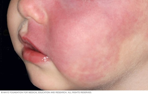
Port-wine stain on an infant’s face
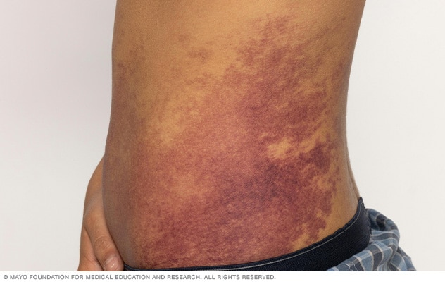
Port-wine birthmark on the side of the torso
Salmon patches
Salmon patches are small areas of skin that may look pink, red or purple. They're often found above the hairline at the back of the neck, on the eyelids or between the eyes. These marks are caused by tiny blood vessels close to the skin. This type of birthmark also is called a stork bite, an angel kiss and a nevus simplex.
Salmon patches may fade as the child grows, though patches at the back of the neck may be permanent. Salmon patches don't need treatment.
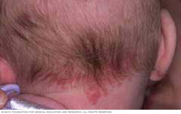
A salmon patch, also called a stork bite, on the back of an infant’s neck.
Hemangioma
A hemangioma is a common birthmark that may look blue, purple or red. It may be present at birth, or it may appear and grow during the first few months after birth. Hemangiomas often appear on the head or neck. They usually get smaller with time. By age 10, many children have only a small mark where the hemangioma was. These birthmarks also are called infantile hemangiomas.
A hemangioma doesn't always need treatment. Medicine or laser treatment may be used if a hemangioma affects a child's appearance or how part of the body works. If a child has several hemangiomas, a healthcare professional may recommend tests to check for hemangiomas inside the body.
Most hemangiomas don't cause serious issues. Seek medical attention if a hemangioma:
- Covers a large area of skin.
- Is located along the middle of the body, especially over the spine.
Depending on their size and location, some hemangiomas can cause complications or be linked to changes inside the body. A healthcare professional can decide whether more testing is needed.
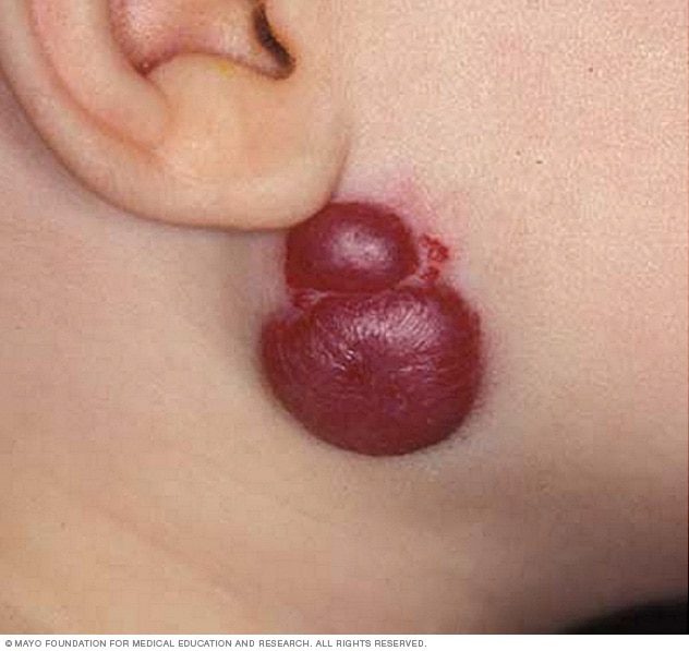
Hemangioma below the ear
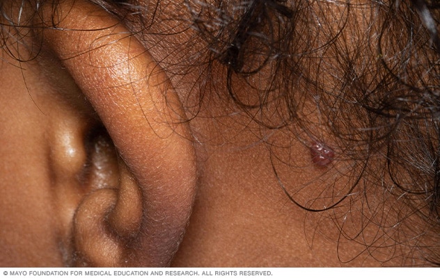
Hemangioma behind the ear
