A fetal ultrasound can be exciting for parents as it offers an early look at a developing baby. But it's also an important tool for the health care team to use to check a fetus's health, including growth and development. It's used to gauge other aspects of pregnancy, too, such as the amount of amniotic fluid and the location of the placenta. In some cases, fetal ultrasound may be used to check for possible problems or to help diagnose a medical issue.
During a fetal ultrasound, a device called a transducer is placed on the pregnant person's belly. Or in some cases, it may be placed in the vagina or on the area between the vagina and the anus. Sound waves are translated into a pattern of light and dark areas. That creates an image of the fetus on a screen, as shown below.
The risks posed by fetal ultrasound are low. But as with all medical procedures, it carries some risk. Fetal ultrasound should only be done for medical reasons as part of prenatal care, based on the advice of a doctor or other licensed health care professional.
If you're getting ready for an ultrasound, check out the following images. They'll give you an idea of what to expect and help you better understand what you see during the ultrasound.
Early pregnancy
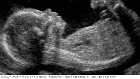
The image below shows a fetus's profile at 11 weeks of pregnancy, which is nine weeks after conception. At this stage, the head makes up about half of a fetus's length.

The heart
The image below shows all four chambers of the heart, as well as the heart valves. This type of image usually is taken during an ultrasound done between weeks 18 and 22 of pregnancy. Fetal ultrasound is used to check that the heart is working properly and to see if there could be any heart problems.

The brain
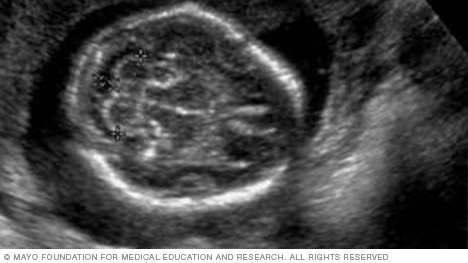
Below is an image of the base of the brain, called the cerebellum. This type of image usually is taken during an ultrasound done between 18 and 22 weeks of pregnancy.
The cerebellum is the part of the brain that controls muscle coordination and balance. Checking its shape on an ultrasound can help the health care team find neural tube defects. The neural tube forms in the first few weeks of pregnancy. The top of the tube becomes the brain. The rest of it becomes the spinal cord. Problems in neural tube development may lead to conditions such as spina bifida, in which part of the neural tube doesn't develop or close correctly. This can lead to problems in the spinal cord and in the bones of the spine.
The head
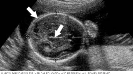
Below is a view of a fetus's head. The thicker white lines that form a circle are the skull. The white line in the middle of the circle is the midline structure that separates the baby's brain into its right and left halves. Head measurements can help determine the age of a fetus.
The hands
Open hand and fingers, as shown below, are among the signs that a health care team looks for on an ultrasound to confirm that a fetus is growing and developing as expected.

The eyes
This image shows the lens of the eye. Twenty-three weeks into pregnancy, which is 21 weeks after conception, rapid eye movements start. The eyelids begin to open at 28 weeks of pregnancy, which is 26 weeks after conception.

The neck
This image is a cross section of the cervical spine. The cervical spine begins at the base of the skull and goes down through the neck. It protects the spinal cord and supports the skull.

The spine
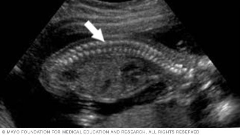
This image is of a fetus's spine. It's one of the easier structures to recognize when viewing a fetal ultrasound.
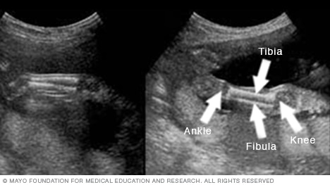
The legs
This image shows the bone in the leg that runs from the hip to the knee, called the femur. It's the largest and strongest bone in the body.

Below are images of the lower legs. The knee is on the right side of each image, and the ankle is on the left. You also can see the shinbone, called the tibia, and the small leg bone on the outside of the ankle, called the fibula.
The umbilical cord
The arrow in the image below points to the site where the umbilical cord is attached to the fetus's belly, also called the abdomen. By looking at this area, the health care team can check for several conditions. They include a condition called omphalocele. It happens when contents of the abdomen come out through an opening at the bellybutton. Another condition is gastroschisis. It involves a break or split in the tissue that forms the abdominal wall.

3D fetal ultrasound
The image below is from a 3D fetal ultrasound. This type of ultrasound can make images that are clearer and more detailed than standard fetal ultrasound images. A 3D fetal ultrasound is sometimes used to detect facial problems, bone problems or neural tube defects.
Some companies offer 3D ultrasounds outside of medical settings. These ultrasounds are often advertised as a way for families to create keepsake images or to learn the sex of a fetus. But most health care professionals advise against getting fetal ultrasounds, including 3D ultrasounds, when there's not a medical need for them.


Mayo Clinic's Ultimate Guide to Pregnancy
This guide offers research-backed advice to help you and your baby experience a healthy pregnancy, written by some of the world's leading medical experts.