Aug. 14, 2026
Highly subspecialized neuroradiologists are linchpins of neurological and neurosurgical care at Mayo Clinic. Advanced imaging technology and clinical training allow Mayo Clinic neuroradiologists across the enterprise to identify conditions that aren't easily seen elsewhere.
"Less than half of all brain MRIs in the United States are read by board-certified neuroradiologists. In patients referred to us, our neuroradiologists have found conditions that may have gone undiagnosed or misdiagnosed for years," says Johnny S. Sandhu, M.D., chair of Neuroradiology at Mayo Clinic in Jacksonville, Florida.
Mayo Clinic uses not only the latest imaging equipment but also sophisticated acquisition techniques. Detailed imaging protocols — often developed by on-site physicists — help capture the previously unseen.
Equally important is expert interpretation of imaging. Mayo Clinic's three main campuses together have more than 80 neuroradiologists. That depth enables a high degree of subspecialization. Just as neurosurgeons might subspecialize in neuro-oncology, the spine or cerebrovascular disease, Mayo Clinic neuroradiologists focus on reading specific types of imaging.
"In patients referred to us, our neuroradiologists have found conditions that may have gone undiagnosed or misdiagnosed for years."
Imaging proficiency paves the way for personalized treatment of the spectrum of neurological and spinal conditions. "As subspecialized neuroradiologists, we can talk the talk with our referring clinical specialists," says Steven A. Messina, M.D., chair of Neuroradiology at Mayo Clinic in Rochester, Minnesota.
"Neuroradiologists play a critical role in the diagnosis and treatment planning of complex neurological and head and neck disorders, and are fully integrated into our care teams," says Tanya J. Rath, M.D., chair of Neuroradiology at Mayo Clinic in Phoenix/Scottsdale, Arizona. "That allows us to provide that next level of expertise for Mayo Clinic patients."
State-of-the-art technology
Mayo Clinic has long been a leader in advanced medical imaging. The enterprise has been an early adopter of some modalities and also develops new technologies in-house.
Current imaging technology includes:
توجيه الرعاية لمرضى تمدد الأوعية
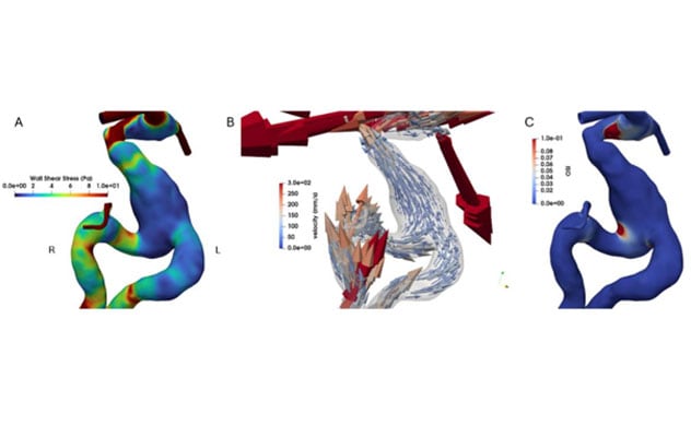
توجيه الرعاية لمرضى تمدد الأوعية
ديناميكا السوائل الحسابية التي ساعدت في تحديد النهج الأمثل لعلاج تمدد الأوعية الدموية المغزلي في الشريان القاعدي. أُعيد إنشاء الصور اعتمادًا على تصوير الأوعية بالرنين المغناطيسي باستخدام تقنية التدفق رباعي الأبعاد.
- 7T MRI, which provides more than twice the resolution of conventional 3T MRI.
- Magnetoencephalography to pinpoint seizure origin locations.
- Photon-counting detector CT, which can provide in-plane resolution of 110 microns for optimal management of several neurological conditions.
- Dynamic susceptibility contrast perfusion MRI to diagnose tumor type and provide additional information for personalized tumor treatment.
- 18F-DOPA PET to identify invasive tumor burden of glioblastoma multiforme.
- Gallium-68-DOTATATE PET to identify specific tumor types and distinguish tumors from nontumoral processes.
- Diffusion tensor imaging for presurgical mapping of white matter tracts to reduce postoperative morbidity and deficits.
- Magnetic resonance (MR) spectroscopy to distinguish tumors from nontumor processes and recurrence from post-treatment effects.
- 4D flow MR angiography, which provides detailed information about blood flow.
- Blood oxygenation level-dependent functional MRI to assess cerebrovascular reserve — sometimes called a brain stress test. This imaging helps determine when cerebrovascular surgery is needed.
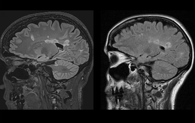 البروتوكول المُحسَّن
على اليسار، تظهر صورة فحص تصويري أُجري في مؤسسة طبية خارجية لمريض مصاب بالتصلّب المتعدد. وعلى اليمين، يَظهر تسلسل T2 FLAIR السهمي للمريض نفسه، وقد أُجري في مايو كلينك باستخدام بروتوكول مُحسَّن. تظهر آفات المادة البيضاء بشكل أوضح وبدقة أعلى في فحص مايو كلينك التصويري.
Imaging protocols are optimized for disease detection. "We incorporate 3D volumetric isotropic imaging that allows us to detect subtle progression in complex diseases," Dr. Messina says.
To further optimize patient care, Mayo Clinic's neuroradiologists work with devicemakers and on-site scientists to develop new imaging protocols. One example is an amyotrophic lateral sclerosis 7T MRI protocol designed by Mayo Clinic physicists and neuroradiologists through extensive volunteer scanning and clinical validation. That imaging can help identify early iron deposition in the motor cortex in patients with upper motor neuron disease. Another example is an in-house 1.5T protocol for postsurgical spinal imaging that significantly decreases artifacts caused by metal implants.
Mayo Clinic also has obtained approval for clinical use of a cerebral vascular reactivity study for patients with intracranial stenosis. Test results can indicate when intervention to prevent stroke is needed. "We are constantly evolving to improve image quality for our patients," Dr. Sandhu says.
Without sacrificing that quality, Mayo Clinic neuroradiologists also strive to shorten the time needed for exams. "We know that the longer the patient is in the scanner, the more likely the images are to be degraded by motion and other potential factors," Dr. Messina says. "We have incredibly high standards for maintaining image quality while mitigating the burden on the patient."
Diagnostic precision
As a quaternary center, Mayo Clinic has technical and clinical resources that aren't always available elsewhere. Those resources can be key to precise diagnosis.
Dr. Sandhu cites a patient referred to Mayo Clinic whose outside MRI showed an area of abnormal signal in the temporal lobe. The MRI finding was interpreted as possibly a low-grade tumor or infection. The lesion was removed at an outside institution, with indeterminate pathology results on the resected tissue.
"But our neuroradiologists, who super-subspecialize in this type of imaging, determined it was a normal tumefactive perivascular space," Dr. Sandhu says. "The patient had been followed with annual MRI to make sure her 'tumor' wasn't growing. After imaging here, we were able to assuage the patient's concern about malignancy and recommended no further surveillance."
The role of neuroradiologists in patient care extends beyond initial diagnosis. They sometimes participate in hospital rounds alongside neurologists and neurosurgeons to guide decision-making about follow-up imaging. "We find that helps avoid unnecessary scans and ensure that clinicians get the answers they need," Dr. Sandhu says.
That team approach epitomizes Mayo Clinic's model of care. "We are very collaborative," Dr. Rath says. "That translates into better care for all Mayo Clinic patients."
For more information
Refer a patient to Mayo Clinic.