March 26, 2022
Mayo Clinic is using radiogenomics to investigate the genetic heterogeneity of individuals' brain tumors. The work — still in its infancy — aims to provide the basis for future therapies that can address multiple genetic targets in a single patient.
"We have some ability right now to tailor treatments to patients. But we need to better understand how each patient can be classified into potential treatment paradigms," says Leland S. Hu, M.D., a neuroradiologist at Mayo Clinic in Phoenix/Scottsdale, Arizona.
توقع الطفرات الوراثية
توقع الطفرات الوراثية
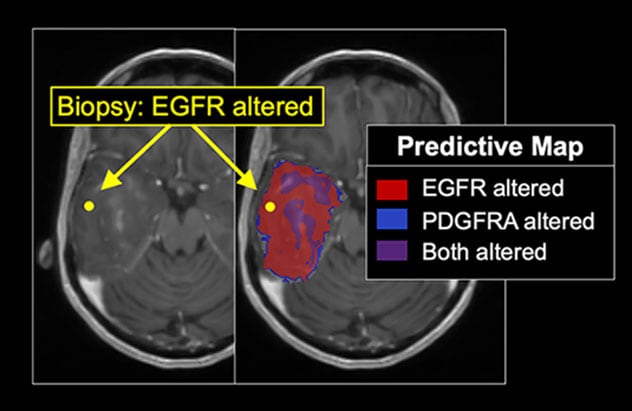
توضح خريطة التصوير الطبي للجينوم مناطق ورم الدماغ التي يتغير فيها مستقبل عامل نمو البشرة ومستقبل عامل النمو المشتق من الصفيحات A وكلاهما. وتوضح الخزعة باستخدام التوجيه التجسيمي (النقطة الصفراء) التوافق مع التوقعات الناحيّة.
Radiogenomics uses machine learning to directly connect the morphological and physiological appearance of tumors on clinical imaging with underlying genomic features. The goal is to use noninvasive MRI to predict the genetic mutations that are present in a tumor — a particular challenge in genetically heterogeneous tumors such as glioblastoma multiforme (GBM).
Mayo Clinic researchers have a unique data set of 95 image-localized biopsies, with spatially matched MRI, from 25 individuals with untreated GBM. The image-localized biopsies provide multiple samples from each tumor.
Applying machine learning to that database, the researchers developed a radiogenomics model to elucidate intratumoral variation. The model — described in the February 2021 issue of Scientific Reports — generated predictions for regional epidermal growth factor receptor (EGFR) amplification at the voxel level throughout different regions of individuals' tumors. EGFR is a key genetic driver of GBM. Unlike similar models, Mayo Clinic's model integrates uncertainty estimates with each spatially resolved model prediction.
An earlier review — published in the May 1, 2020, issue of Cancer Letters — outlined the clinical applications of advanced neuro-oncologic imaging for the assessment of intratumoral heterogeneity.
"We're using imaging to give us a sense of the tumor's biology at the micro level. We're already seeing some differences in the genetic drug targets from a single tumor," Dr. Hu says. "We'd like to look at the individual pixel level and predict the genetic drug targets within that pixel — and then extend that analysis over the whole tumor, to give us a broad overview of where the genetic drug targets are located."
Models to determine tumor extent and recurrence
Mayo Clinic researchers are also using radiomics to investigate certain nongenetic aspects of tumor biology. One project aims to better determine tumor extent. As with the radiogenomics work, machine learning is applied to MRI and image-localized biopsy data.
"That allows us to build spatially accurate models that can tell us the extent of tumor beyond what we can see from conventional T1-weighted post-gadolinium MRI," Dr. Hu says. "The goal is to potentially guide more-extensive surgical resection or more-extended, higher dose radiation therapy in areas of tumor that aren't visible in conventional MRI."
Other work involves the use of dynamic susceptibility contrast magnetic resonance perfusion to differentiate recurrent tumor from treatment effect in individuals with high-grade gliomas. In a study published in the March 2020 edition of the American Journal of Neuroradiology, Mayo Clinic researchers found that standardization of relative cerebral blood volume measurements can provide that distinction. That Mayo Clinic paper received the journal's Lucien Levy Best Research Article Award in 2019.
"Much of the challenge in differentiating recurrent tumor from treatment effect is that the treatment bed can have a mixture of both," Dr. Hu says. "Sampling different regions of a lesion gives us better spatial resolution to distinguish what parts are tumor and what parts are post-treatment enhancement."
Dr. Hu notes that translating radiogenomic and radiomic research into targeted clinical therapies will require years of further work. To reach that goal, Mayo Clinic deploys a multidisciplinary team including neuroradiologists, neuro-oncologists, cancer biologists, imaging physicists and experts in machine learning and mathematical modeling.
"It's a huge team effort with a very unified perspective," Dr. Hu says. "We have a common goal of advancing this field to help patients."
For more information
Hu LS, et al. Uncertainty quantification in the radiogenomics modeling of EGFR amplification in glioblastoma. Scientific Reports. 2021;11:3932.
Hu LS, et al. Imaging of intratumoral heterogeneity in high-grade glioma. Cancer Letters. 2020;427:97.
Hoxworth JM, et al. Performance of standardized relative CBV for quantifying regional histologic tumor burden in recurrent high-grade glioma: Comparison against normalized relative CBV using image-localized stereotactic biopsies. American Journal of Neuroradiology. 2020;41:408.
Refer a patient to Mayo Clinic.