May 21, 2021
فهم التشريح
فهم التشريح
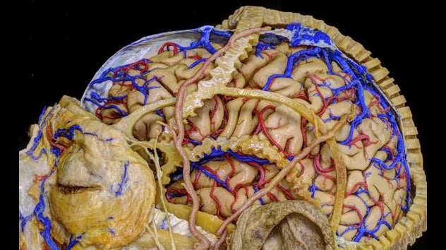
يساعد التشريح المجهري المفصل للغاية لعينة دماغ جُثية في بناء المعرفة بتشريح الدماغ.
فهم التشريح
فهم التشريح
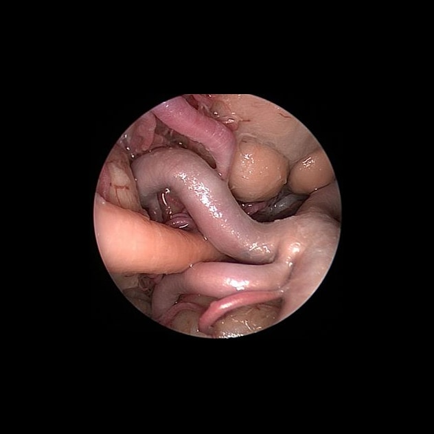
يساعد التشريح التنظيري المفصل للغاية لعينة دماغ جُثية في بناء المعرفة بتشريح الدماغ.
The treatment of complex skull base pathology requires careful surgical navigation through patient anatomy that is distorted by disease. Building on efforts begun more than a half-century ago, Mayo Clinic's clinician-researchers are working to learn more about cranial anatomy and skull base disease to enhance the safety and outcome of surgical treatment.
"Understanding how pathology affects delicate structures such as intracranial nerves and blood vessels and discovering new ways to reach the pathology safely are very important for the success of skull base surgery," says Maria Peris Celda, M.D., Ph.D., a neurosurgeon specializing in complex cranial conditions at Mayo Clinic in Rochester, Minnesota.
In addition to managing her surgical practice, Dr. Peris Celda leads a laboratory team that is studying clinical features of complex skull base tumors as well as working to develop and validate new surgical techniques. The laboratory provides a training environment for residents and fellows from around the world.
This combination of clinical practice, research and education extends the legacy of the late Albert L. Rhoton Jr., M.D., a world-renowned neurosurgeon and a pioneer in the study of microneurosurgical anatomy in neurosurgery who had his practice at Mayo Clinic in Rochester from 1966 to 1972. Dr. Peris Celda completed her surgical anatomy fellowship under Dr. Rhoton in 2013 at the University of Florida.
"There's never a finish line to the study of surgical anatomy," Dr. Peris Celda says. "We are constantly developing new techniques and approaches that must be validated in the anatomy laboratory."
Patient-specific care
Mayo Clinic takes a multidisciplinary approach to complex intracranial disease. In addition to neurosurgeons, there are specialists in Radiology, Otolaryngology (ENT)/Head and Neck Surgery, Ophthalmology, Neurology, Oncology, Radiation Oncology, and Pathology on the treatment team.
Subspecialized neuroradiologic expertise is a key aspect of care. Mayo Clinic uses advanced imaging to guide surgical planning.
"New MRI techniques and 3D printing technologies based on imaging are especially useful in allowing us to evaluate the tumor's consistency and the anatomical relationships that are essential to perform safe operations," Dr. Peris Celda says. "Our laboratory has an ongoing collaboration with Mayo's 3D modeling laboratory to create patient-specific surgical models and to better characterize important clinical features of skull base tumors."
For patients needing surgery, decisions about optimal approaches are based on the individuals' needs. Mayo Clinic surgeons perform minimally invasive, open or endoscopic procedures, or sometimes a combination of these procedures.
"We are working toward greater understanding of complex skull base diseases and the ability to predict the anatomy that we will find in surgery" Dr. Peris Celda says. "With the aid of surgical anatomy knowledge, new MRI techniques and 3D modeling, we will be able to innovate the neurosurgical field toward safer, more effective and minimally invasive surgeries."
For more information
3D Anatomic Modeling Laboratories. Mayo Clinic.