Diagnosis
Ductal carcinoma in situ (DCIS) is most often discovered during a mammogram used to screen for breast cancer. A mammogram is an X-ray of the breast tissue. If your mammogram shows something concerning, you will likely have additional breast imaging and a biopsy.
Mammogram
Breast calcifications
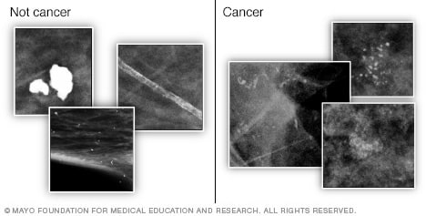
Breast calcifications
Calcifications are small buildups of calcium, called calcium deposits, in the breast. They show up as white spots on a mammogram. Large, round or well-defined calcifications (shown left) are more likely to not be cancer, also called benign. Tight clusters of tiny, fine, irregularly shaped calcifications (shown right) may mean cancer.
If an area of concern was found during a screening mammogram, you may then have a diagnostic mammogram. A diagnostic mammogram takes views at higher magnification from more angles than a mammogram used for screening. This examination evaluates both breasts.
A diagnostic mammogram gives your healthcare team a closer look at any calcium deposits detected in the breast tissue. Calcium deposits, also called calcifications, can sometimes be cancerous.
With DCIS, the calcifications are often irregularly shaped, which mammogram results might describe as pleomorphic. The calcifications often are grouped together. Sometimes the calcifications look like thin lines on mammogram images.
If the area of concern needs further evaluation, the next step may be an ultrasound and a breast biopsy.
Breast ultrasound
Ultrasound uses sound waves to make images of structures inside the body. A breast ultrasound may give your healthcare team more information about an area of concern. The healthcare team uses this information to decide what tests you might need next.
Breast MRI
Breast MRI
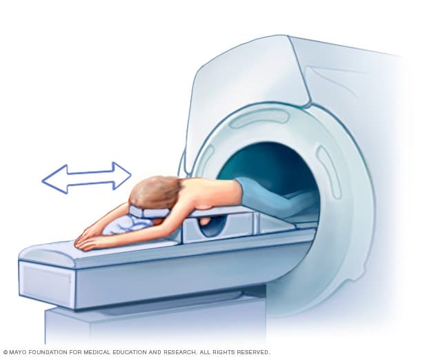
Breast MRI
Getting a breast MRI involves lying face down on a padded scanning table. The breasts fit into a hollow space in the table. The hollow has coils that get signals from the MRI. The table slides into the large opening of the MRI machine.
MRI machines use a magnetic field and radio waves to create pictures of the inside of the body. A breast MRI can make more-detailed pictures of the breast. Before a breast MRI, you usually receive an injection of contrast. The contrast material helps the tissue show up better in the images.
Biopsy
Stereotactic breast biopsy
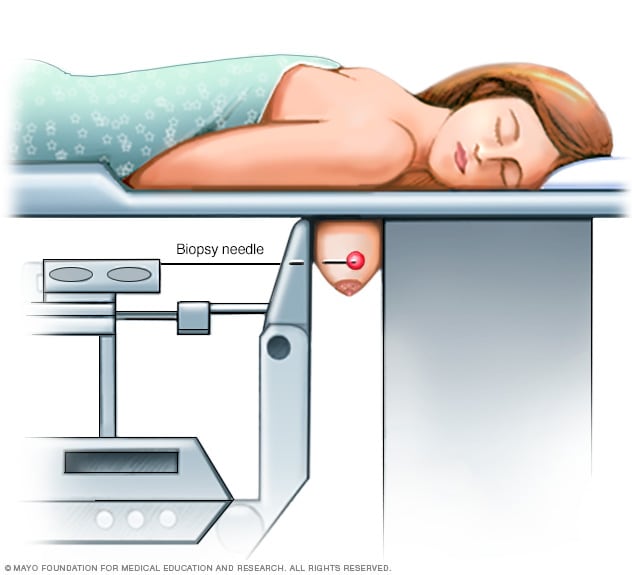
Stereotactic breast biopsy
During a stereotactic breast biopsy, the breast is firmly compressed between two plates. Breast X-rays, called mammograms, are used to produce stereo images. Stereo images are images of the same area from different angles. They help find the exact location for the biopsy. A sample of breast tissue from the suspicious area is then removed with a needle.
A biopsy is a procedure to remove a sample of tissue for testing in a lab. For a breast biopsy, a healthcare professional puts a needle through the skin and into the breast tissue. Imaging, such as X-rays or ultrasound, helps guide the needle to the right place. The healthcare professional uses the needle to draw out tissue from the breast. A marker may be placed in the spot where the tissue sample was removed. This small metal marker shows up on imaging tests. The marker helps your care team find the area of concern.
Lab testing
The tissue sample from a biopsy goes to a lab for testing. Tests can show whether the cells in the sample are cancerous. Other tests give more details about the cells. The testing is done by pathologists. Pathologists are doctors who look for disease in cells, fluids and tissue removed from the body.
Results are often given in a pathology report. The results may show:
- DCIS pattern. DCIS can be divided into types that describe how the cells grow within a breast duct. These patterns of growth include comedo, cribriform, micropapillary, papillary and solid.
- Cell grade. The grade of the cancer cells is based on how the cells look under a microscope. The grade tells the healthcare team whether the cancer is likely to grow slowly or quickly.
- Hormone receptor status. Most breast cancer cells use the hormones estrogen or progesterone to help them grow. Cells that do this have receptors that help them catch the hormones circulating in the bloodstream. If testing finds receptors for one or both hormones, the cancer is hormone receptor positive. If testing doesn't find receptors, then the cancer is hormone receptor negative.
- Necrosis. DCIS sometimes has areas of dead cells among the cancer cells. Healthcare professionals call these areas necrosis. Small areas of necrosis are called focal necrosis. Larger areas of necrosis in the center of the cancer are called comedo necrosis.
DCIS usually isn't tested for the presence of a protein called HER2. Invasive breast cancers are tested for HER2, but noninvasive cancers generally aren't.
More Information
Grades
A cancer's grade is a number that tells the healthcare team how different the cancer cells look from healthy cells. The grades of ductal carcinoma in situ (DCIS) go from 1 to 3. The grade helps the healthcare team predict how the DCIS may behave over time.
To decide on the grade, doctors in the lab, called pathologists, use a microscope to look at the cancer cells. If the cancer cells look similar to healthy cells, then the cancer cells are low grade. Low-grade cancer grows slowly. If the cancer cells look very different from healthy cells, then the cancer cells are high grade. High-grade cancer grows quickly.
The grades of DCIS include:
- Grade 1. Grade 1 is a low-grade DCIS. The cancer cells look similar to healthy breast cells. Grade 1 DCIS has a lower risk of returning after treatment or becoming invasive.
- Grade 2. Grade 2 is an intermediate-grade DCIS. The cancer cells look somewhat different from healthy breast cells. Grade 2 DCIS has an intermediate risk of returning after treatment or becoming invasive.
- Grade 3. Grade 3 is a high-grade DCIS. The cancer cells look very different from healthy breast cells. Grade 3 DCIS has the highest risk of returning after treatment or becoming invasive.
The grade helps the healthcare team understand the prognosis and make a treatment plan. High-grade DCIS is more likely to return after surgery, so additional treatment may be recommended. Lower grade DCIS has a lower risk of recurrence, so additional treatment may not always be needed.
Treatment
Ductal carcinoma in situ (DCIS) is usually treated with surgery to remove the cancer. Sometimes other treatments are used after surgery to lower the risk that the cancer will come back. These treatments may include radiation therapy after lumpectomy surgery and estrogen blocker therapy if the DCIS is hormone receptor positive.
DCIS treatment has a high likelihood of success, and this cancer often can be cured. After treatment, the chance that DCIS will come back or become invasive is usually low.
Healthcare professionals can't always predict which cancers are more likely to come back after treatment. For this reason, they may recommend additional treatment after surgery to lower that risk. This is called adjuvant therapy.
Deciding whether to have adjuvant therapy often involves talking with your healthcare team about the possible benefits and risks. Your decision may depend on your specific situation and your preferences.
In most people, treatment options for DCIS include:
- Breast-conserving surgery, called a lumpectomy, and radiation therapy.
- Breast-removing surgery, called a mastectomy.
In some people, treatment options may include:
- Lumpectomy only.
- Lumpectomy and estrogen blocker therapy.
Surgery
Lumpectomy
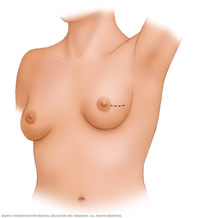
Lumpectomy
A lumpectomy involves removing the cancer and some of the healthy tissue that surrounds it. This illustration shows one possible incision that can be used for this procedure, though your surgeon will determine the approach that's best for your particular situation.
If you're diagnosed with DCIS, one of the first decisions you'll have to make is whether to treat the condition with lumpectomy or mastectomy.
-
Lumpectomy. A lumpectomy is surgery to remove the breast cancer and some of the healthy tissue around it. It allows you to maintain most of your breast. Other names for this surgery are breast-conserving surgery and wide local excision. Most people have radiation therapy after lumpectomy.
Research suggests that there is a slightly higher risk of the cancer coming back after lumpectomy compared with mastectomy. However, survival rates between the two treatment approaches are very similar.
If you have other serious health conditions, you might consider other options, such as lumpectomy plus hormone therapy, lumpectomy alone or no treatment.
- Mastectomy. A mastectomy is surgery to remove all breast tissue from a breast. Total mastectomy also is called simple mastectomy. It removes all of the breast, including the lobules, ducts, fatty tissue and some skin, as well as the nipple and areola. Other procedures include skin-sparing mastectomy, which leaves the skin, and nipple-sparing mastectomy, which leaves the areola, nipple and skin. Breast reconstruction to restore the appearance of the breast can be done at the same time or in a later procedure, if you desire.
Lumpectomy is a good option for most people with DCIS. But mastectomy may be recommended if:
- You have a large area of DCIS. If the area is large relative to the size of your breast, a lumpectomy may not produce acceptable cosmetic results.
- There's more than one area of DCIS. When multiple areas are involved, it is called multifocal or multicentric disease. It's difficult to remove multiple areas of DCIS with a lumpectomy. This is especially true if the areas are found in different parts of the breast.
- Biopsy results show cancer cells at or near the edge of the tissue sample. There may be more DCIS than originally thought. This means that a lumpectomy might not be enough to remove all affected areas. A mastectomy could be needed to remove all of the breast tissue.
- You're not a candidate for radiation therapy. Radiation is usually given after a lumpectomy. Radiation might not be an option if you're in the first trimester of pregnancy or if you've received radiation to your chest or breast in the past. It also might not be recommended if you have a condition that makes you more sensitive to radiation side effects, such as systemic lupus erythematosus.
- You prefer to have a mastectomy. For instance, you might not want a lumpectomy if you don't want to have radiation therapy.
Because DCIS is noninvasive, surgery typically doesn't involve the removal of lymph nodes from under your arm. The chance of finding cancer in the lymph nodes is extremely small.
If the cancer cells may have spread outside the breast duct or if you are having a mastectomy, then some lymph nodes may be removed as part of the surgery.
Radiation therapy
Radiation therapy
Radiation therapy
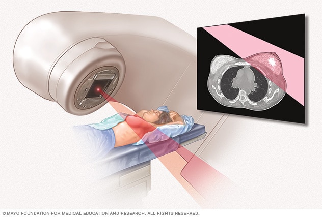
External beam radiation uses high-powered beams of energy to kill cancer cells. Beams of radiation are precisely aimed at the cancer using a machine that moves around your body.
Radiation therapy treats cancer with powerful energy beams. The energy can come from X-rays, protons or other sources.
Radiation therapy for breast cancer often uses external beam radiation. During this type of radiation therapy, you lie on a table while a machine moves around you. The machine directs radiation to precise points on your body.
For DCIS, radiation therapy is often used after lumpectomy to reduce the chance that the cancer will come back. But it might not be necessary if you have only a small area of DCIS that is considered slow growing and was completely removed during surgery.
Radiation can be applied to the entire breast, called whole-breast radiation, or to part of the breast, called partial-breast radiation. Your healthcare team may talk about these options with you after surgery. The approach your team recommends may depend on your age and the size, grade and hormone receptor status of your cancer.
Estrogen blocker therapy
Estrogen blocker therapy treats breast cancer that is sensitive to hormones. This type of cancer has cells with proteins called receptors. The receptors attach to the hormones estrogen and progesterone. The cancer grows in response to these hormones. Healthcare professionals call these cancers hormone receptor positive. DCIS is usually sensitive to hormones.
For DCIS, estrogen blocker therapy is typically used after surgery or radiation. It lowers the risk that the cancer will come back. It also reduces the risk of developing another breast cancer.
Estrogen blocker therapy treatments can include:
- Medicines that block or destroy hormone receptors. These medicines are called selective estrogen receptor modulators. This category includes the medicine tamoxifen. It may be used before or after menopause.
- Medicines that lower hormone production. These medicines are called aromatase inhibitors. They may be used only after menopause.
Discuss the benefits and risks of estrogen blocker therapy with your healthcare team.
More Information
Clinical trials
Explore Mayo Clinic studies testing new treatments, interventions and tests as a means to prevent, detect, treat or manage this condition.
Alternative medicine
No alternative medicine treatments have been found to cure ductal carcinoma in situ (DCIS). But complementary and alternative medicine therapies may help you cope with side effects of treatment.
Combined with your healthcare team's recommendations, complementary and alternative medicine treatments may provide some comfort. Examples include:
- Art therapy.
- Exercise.
- Meditation.
- Music therapy.
- Relaxation exercises.
- Spirituality.
Coping and support
A diagnosis of ductal carcinoma in situ (DCIS) can be unexpected, and it's natural to have questions about what comes next. You may be receiving a lot of information and facing decisions about surgery, radiation and medical treatments. It's common to have a range of feelings that may change from day to day, and it can take time to process everything. As you move forward, here are some ideas that might be helpful.
Learn enough about DCIS to make decisions about your care
Ask your healthcare team questions about your diagnosis and your pathology results. Use this information to research your treatment options.
Knowing more about your cancer and your options may help you feel more confident when making treatment decisions. Still, some people don't want to know the details of their cancer. If this is how you feel, let your care team know that too.
Find a good listener
Finding someone who is willing to listen to you talk about your hopes and fears can be helpful as you manage a cancer diagnosis. This could be a friend or family member. A counselor, medical social worker or clergy member also may offer helpful guidance and care.
Talk with other breast cancer survivors
You may find it helpful and encouraging to talk to others who have been diagnosed with breast cancer. Ask your healthcare team about support groups in your area, or contact the American Cancer Society or another cancer organization in your area to find resources. Find support online through Mayo Clinic Connect, a community where you can connect with others for support, practical information and answers to everyday questions.
Preparing for your appointment
Make an appointment with a doctor or other healthcare professional if you have any symptoms that worry you. If an exam or imaging test shows that you might have ductal carcinoma in situ (DCIS), your healthcare team will likely refer you to a specialist.
Specialists who care for people with DCIS include:
- Breast medicine specialists.
- Breast surgeons.
- Genetic counselors.
- Oncologists. These doctors specialize in treating cancer.
- Plastic surgeons.
- Radiation oncologists. These doctors treat cancer with radiation.
- Radiologists. These doctors specialize in diagnostic tests, such as mammograms.
Here's some information to help you get ready for your appointment.
What you can do
- Write down your medical history, including any benign breast conditions with which you've been diagnosed. Also mention any radiation therapy you may have received, even years ago.
- Write down your family history of cancer. Note any family members who have had cancer. Note how each member is related to you, the type of cancer, the age at diagnosis and whether each person survived.
- Make a list of all medicines, vitamins or supplements that you're taking. If you are currently taking or have previously taken hormone replacement therapy, tell your healthcare professional.
- Consider taking a family member or friend along. Sometimes it can be difficult to take in all the information you receive during an appointment. Someone who goes with you may remember something that you missed or forgot.
- Write down questions to ask your healthcare professional.
Questions to ask your doctor
Your time with your healthcare professional is limited. Prepare a list of questions so that you can make the most of your time together. List your questions from most important to least important in case time runs out. For breast cancer, some basic questions to ask include:
- Do I have breast cancer?
- What tests do I need to determine the type and stage of cancer?
- What treatment approach do you recommend?
- What are the possible side effects or complications of this treatment?
- In general, how effective is this treatment?
- Am I at risk of this condition recurring?
- Am I at risk of developing invasive breast cancer?
- How will you treat DCIS if it returns?
- How often will I need follow-up visits after I finish treatment?
- What lifestyle changes can help reduce my risk of a DCIS recurrence?
- Do I need a second opinion?
- Should I see a genetic counselor?
In addition to the questions that you've prepared, don't hesitate to ask other questions you think of during your appointment.
What to expect from your doctor
Be prepared to answer some questions about your symptoms and your health, such as:
- Have you gone through menopause?
- Are you using or have you used any medicines or supplements to relieve the symptoms of menopause?
- Have you had other breast biopsies or operations?
- Have you been diagnosed with any breast conditions, including noncancerous conditions?
- Have you been diagnosed with any other medical conditions?
- Do you have any family history of breast cancer?
- Have you or your blood relatives ever been tested for BRCA gene changes?
- Have you ever had radiation therapy?
- What is your typical daily diet, including alcohol intake?
- Are you physically active?
Aug. 08, 2026
- Niederhuber JE, et al., eds. Cancer of the breast. In: Abeloff's Clinical Oncology. 6th ed. Elsevier; 2020. https://www.clinicalkey.com. Accessed March 1, 2024.
- Townsend CM Jr, et al. Diseases of the breast. In: Sabiston Textbook of Surgery: The Biological Basis of Modern Surgical Practice. 21st ed. Elsevier; 2022. https://www.clinicalkey.com. Accessed March 1, 2024.
- Breast cancer. National Comprehensive Cancer Network. https://www.nccn.org/guidelines/guidelines-detail?category=1&id=1419. Accessed June 1, 2026.
- Abraham J, et al., eds. Breast cancer. In: The Bethesda Handbook of Clinical Oncology. 6th ed. Kindle edition. Wolters Kluwer; 2023. Accessed March 1, 2024.
- Collins LC, et al. Breast ductal carcinoma in situ: Epidemiology, clinical manifestations, and diagnosis. https://www.uptodate.com/contents/search. Accessed June 3, 2026.
- Distress management. National Comprehensive Cancer Network. https://www.nccn.org/guidelines/guidelines-detail?category=3&id=1431. Accessed March 1, 2024.
- Record SM, et al. How to navigate the treatment spectrum from multimodality therapy to observation alone for ductal carcinoma in situ. Surgical Oncology Clinics of North America. 2023; doi:10.1016/j.soc.2023.05.011.
- Breast cancer prevention (PDQ) – Patient version. National Cancer Institute. https://www.cancer.gov/types/breast/patient/breast-prevention-pdq. Accessed March 1, 2024.
- In situ. Dorland's Medical Dictionary Online. https://www.dorlandsonline.com. Accessed June 5, 2026.
- Klimberg VS, et al., eds. Bland and Copeland's The Breast: Comprehensive Management of Benign and Malignant Diseases. 6th ed. Elsevier; 2024. https://www.clinicalkey.com. Accessed June 3, 2026.
- Collins LC, et al. Ductal carcinoma in situ: Treatment and prognosis. https://www.uptodate.com/contents/search. Accessed June 1, 2026.
- Collins LC, et al. Microinvasive breast carcinoma. https://www.uptodate.com/contents/search. Accessed June 3, 2026.
- Bleiweiss IJ. Pathology of breast cancer. https://www.uptodate.com/contents/search. Accessed June 3, 2026.
- Dabbs DJ. Ductal carcinoma in situ. In: Breast Pathology. 3rd ed. Elsevier; 2024. https://www.clinicalkey.com. Accessed June 5, 2026.
- Protocol for the examination of biopsy specimens from patients with ductal carcinoma in situ (DCIS) of the breast. College of American Pathologists. https://www.cap.org/protocols-and-guidelines/cancer-reporting-tools/cancer-protocol-templates. Accessed June 4, 2026.
- Cribriform. Dorland's Medical Dictionary Online. https://www.dorlandsonline.com. Accessed June 5, 2026.
- Delaloge S, et al. Ductal carcinoma in situ of the breast: Finding the balance between overtreatment and undertreatment. The Lancet. 2024; doi:10.1016/S0140-6736(24)00425-2.
- Protocol for the examination of resection specimens from patients with ductal carcinoma in situ (DCIS) of the breast. College of American Pathologists. https://www.cap.org/protocols-and-guidelines/cancer-reporting-tools/cancer-protocol-templates. Accessed June 4, 2026.
- Tumor grade. National Cancer Institute. https://www.cancer.gov/about-cancer/diagnosis-staging/diagnosis/tumor-grade. Accessed April 16, 2026.
- Worni M, et al. Trends in treatment patterns and outcomes for ductal carcinoma in situ. Journal of the National Cancer Institute. 2015; doi:10.1093/jnci/djv263.
- Breast SPOREs. National Cancer Institute. https://dctd.cancer.gov/research/spores/focus-area/breast. Accessed April 17, 2026.
- Member institutions. Alliance for Clinical Trials in Oncology. https://www.allianceforclinicaltrialsinoncology.org/main/public/standard.xhtml?path=%2FPublic%2FInstitutions. Accessed April 17, 2026.
- Andreas A, et al., eds. The breast. In: Grainger & Allison's Diagnostic Radiology: A Textbook of Medical Imaging. 7th ed. Elsevier; 2021. https://www.clinicalkey.com. Accessed July 1, 2026.
- AskMayoExpert. Nipple discharge. Mayo Clinic; 2024.
Related
Associated Procedures
Products & Services
Ductal carcinoma in situ (DCIS)