Overview
Cerebral cavernous malformation
Cerebral cavernous malformation
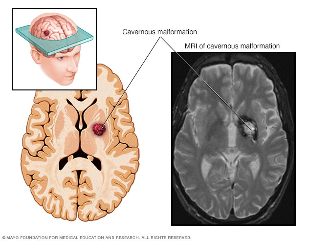
A cerebral cavernous malformation is an irregularly formed blood vessel, shaped like a small mulberry. It can form in the brain or spinal cord and may result in a wide range of neurological symptoms.
Cerebral cavernous malformations (CCMs) are small clusters of blood vessels in the brain or spinal cord. The vessels contain slow-moving blood that's usually clotted. CCMs look like small mulberries and have thin walls that may leak blood.
Cavernous malformations vary in size. Often they are less than 1/2 inch (1 centimeter). Most CCMs are sporadic. This means the CCM happens by chance. It shows up by itself in someone who has no family history of the condition. But about 20% of CCMs are familial. These run in families. Familial CCMs are passed down because of a change in genes. People with familial CCMs usually have more than one cavernous malformation.
A CCM is one of several types of brain vascular malformations that contain irregular blood vessels. Other types of vascular malformations include:
- Arteriovenous malformation (AVM).
- Dural arteriovenous fistula.
- Developmental venous anomaly (DVA).
- Capillary telangiectasia.
For people who have the sporadic form, it's common to have both a DVA and a CCM.
CCMs may leak blood and lead to bleeding in the brain or spinal cord, known as a hemorrhage. Brain hemorrhages can cause many symptoms, such as seizures.
Depending on the location, CCMs also can cause strokelike symptoms such as trouble with movement or feeling in the legs and sometimes the arms. CCMs also may cause bowel and bladder symptoms.
Products & Services
Symptoms
Most cerebral cavernous malformations (CCMs) do not cause symptoms. When symptoms occur, they can vary based on where the CCM is located:
- If a CCM is in the brain, it can sometimes lead to seizures or other bleeding-related symptoms such as weakness, trouble speaking or changes in vision.
- If a CCM is in the spinal cord, the symptoms can vary. For example, a CCM in your spinal cord may cause trouble with your bowel and bladder or make it hard to move or feel your legs or arms.
Generally, symptoms of CCMs may include:
- Seizures.
- A stroke caused by bleeding in the brain, called a hemorrhagic stroke.
- Bad headaches.
- Weakness in the arms or legs.
- Numbness.
- Trouble speaking.
- Poor memory and attention.
- Trouble balancing and walking.
- Vision changes, such as double vision.
Symptoms can get worse over time with repeated bleeding. Bleeding can happen again soon after the first bleed or much later. In some people, a repeat bleed may never occur.
When to see a doctor
Seek medical help right away if you have any symptoms of a seizure. Also get medical help right away if you have symptoms that suggest a cerebral cavernous malformation or brain bleeding.
Causes
Most cerebral cavernous malformations (CCMs) happen by chance. This is called the sporadic form. It means there is only one malformation without any family history. The sporadic form often happens with another irregular vein that looks like a witch's broom. This is called a developmental venous anomaly (DVA).
However, about 20% of people with a CCM have a genetic form. This form is passed down in families, known as familial cavernous malformation syndrome. People with this form may have family members with CCMs, most often with more than one malformation. A diagnosis can be confirmed by a genetic test that requires a blood or saliva sample. Genetic testing is often recommended for people who have:
- MRI evidence of multiple CCMs without a DVA.
- A family history of CCMs.
Radiation to the brain or spinal cord also may result in CCMs within 2 to 20 years afterward. Other rare syndromes may be associated with CCM.
Risk factors
Most cerebral cavernous malformations (CCMs) have no clear cause. But the form that's passed down through families can cause multiple CCMs, both to start with and over time.
To date, research has identified three genetic changes responsible for cavernous malformations passed down through families. Almost all familial cases of cavernous malformations have been traced through those genetic changes.
Familial CCMs are passed down in families through a change in one of these genes:
- KRIT1, also called CCM1.
- MGC4607, also called CCM2.
- PDCD10, also called CCM3.
These genes are responsible for affecting the leakiness of blood vessels and the proteins that keep the blood vessel cells together.
Familial CCMs change over time. They may increase in number or increase or decrease in size. The most serious form of the disease is usually seen in people who have changes in the CCM3 gene. People with CCM3 changes may have symptoms at a younger age, more lesions and a greater risk of hemorrhage.
Complications
The most serious complications of cerebral cavernous malformations (CCMs) stem from repeated bleeding, known as hemorrhages. CCMs that bleed over and over again may cause a hemorrhagic stroke and lead to damage in the nervous system.
Bleeding is more likely to return in people with prior hemorrhages. Bleeding also is more likely to happen again with CCMs located in the brainstem.
Prevention
Cerebral cavernous malformations (CCMs) can't be prevented. This is because the exact cause of CCMs isn't known. It's also because the genes that cause familial CCMs can be passed down through families through genes.
Products & Services