Aug. 29, 2025
Mayo Clinic manufactures 3D models of patient anatomy and pathology to guide complex neurosurgical care. Created in house, the life-size, personalized models give neurosurgeons a tangible understanding of a patient's condition that is often difficult to fully grasp through traditional imaging alone.
 无需进行思维体操
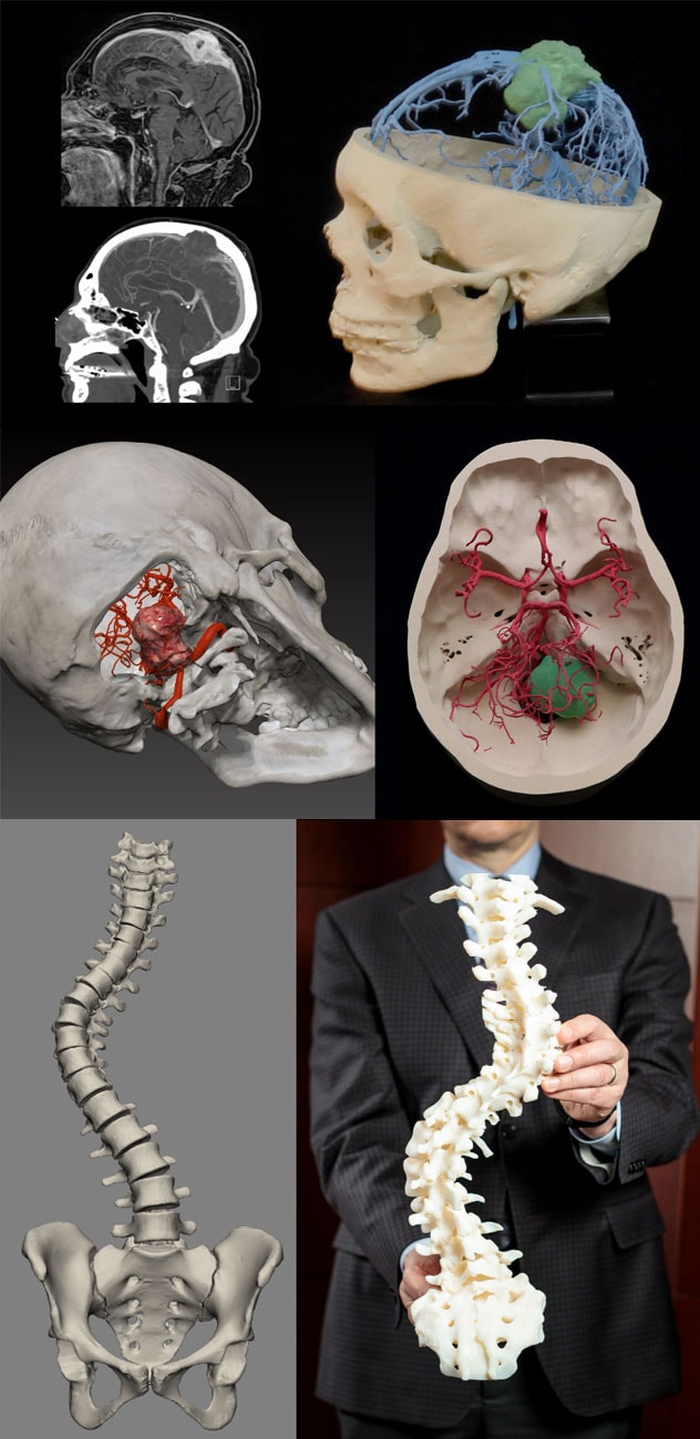
妙佑医疗国际制作的真人大小、针对特定患者的三维解剖模型展示了复杂的颅内肿瘤(上图)、颅底肿瘤(中图)和脊柱弯曲(下图)。
"Modern-day CT and MRI scans can generate over 15,000 images for surgeons to view and try to make a patient-specific 3D model in their heads. The digital twins we manufacture can be held in the surgeons' hands, taking away those mental gymnastics," says Jonathan M. Morris, M.D., a neuroradiologist at Mayo Clinic in Rochester, Minnesota, and medical director of Mayo Clinic's 3D Anatomic Modeling Laboratories.
The anatomic modeling unit (AMU) at Mayo Clinic's campus in Minnesota is the largest point-of-care additive manufacturing facility in the Western Hemisphere. Mayo Clinic in Jacksonville, Florida, and Mayo Clinic in Phoenix/Scottsdale, Arizona, also have 3D anatomic modeling laboratories, providing an enterprise-wide approach to manufacturing. The three laboratories serve all medical and surgical specialties. In neurology and neurosurgery, the models are especially helpful when patients need treatment for congenital spinal anomalies, craniofacial reconstructions, or skull base, brain or spinal tumors.
"Being able to feel in real size what a patient's tumor and anatomy look like has really changed my practice," says Maria Peris Celda, M.D., Ph.D., a neurosurgeon specializing in skull base tumors at Mayo Clinic's campus in Minnesota. "The models are important for deciding the best approach for tumors that are difficult to access. 3D anatomic modeling is helping to advance neurosurgery."
At Mayo Clinic, 3D anatomic modeling and custom medical device manufacturing have scaled to become seamlessly integrated into care delivery. "We've created an infrastructure that allows any clinician in our enterprise to press a button in the electronic medical record and order an anatomical model, custom cutting guide, virtual surgical planning or a custom medical device," Dr. Morris says. "Ultimately, it's about providing the best possible outcomes for our patients who come with complex medical problems."
Meeting patients' needs, in-house
Mayo Clinic's unique approach to anatomic modeling began with an effort to meet the needs of two specific patients. In 2004, a team of Mayo Clinic specialists successfully separated twins conjoined at the chest. While planning the challenging procedure, one of the surgeons asked Dr. Morris and colleagues for a 3D model of the babies' liver. "We outsourced the project, and it came back not looking like a liver," Dr. Morris says.
The physicians then turned to Mayo Clinic's engineering division, whose engineers were able to create the needed 3D model in-house. The AMU grew from there.
"Being able to feel in real size what a patient's tumor and anatomy look like has really changed my practice."
The manufacturing system is a multidisciplinary effort involving radiologists, surgeons, engineers and healthcare technicians. At Mayo Clinic's campus in Minnesota, the AMU and engineering division together have 47 3D printers that can print with several plastics, biochemical resins and titanium. About 20 printers are located above the operating room in Mayo Clinic Hospital, Saint Marys Campus.
"That's where most of our surgeons practice. They can come up to the lab for 10 minutes if they need to discuss something between cases," Dr. Morris says. "Working with an outside vendor, we'd have to schedule a one-hour online meeting, then sometimes a second meeting. Our manufacturing ecosystem fits into a busy clinical infrastructure, starting with an order in the electronic medical record and ultimately a delivery to the surgeon."
The manufacturing process begins with advanced CT or MRI scans of individual patients. Radiologists use sophisticated software to process the scans to create virtual 3D models. One or more printer technologies are used to print the 3D models. Sterilizable models and devices can be created, for use in the operating room or implantation in patients. Postprinting processing readies the models for use, with a robust quality control system providing accuracy and safety for patients. The manufacturing process follows national ASTM standards.
"We have quality control checks and validation every step along the way," Dr. Morris says. "Our system ensures that the patient-specific 3D model we make is exactly what a surgeon will find in the OR."
For neurosurgeons, that knowledge is key to guiding challenging procedures, such as complex scoliosis correction. "The 3D model allows us to see critical structures that traditional 2D imaging doesn't show. That gives us the comfort level to be more aggressive in the corrections we undertake," says Kingsley Abode-Iyamah, M.D., a spinal neurosurgeon at Mayo Clinic's campus in Florida.
He cites a 22-year-old woman with neurofibromatosis who came to Mayo Clinic after multiple failed scoliosis surgeries elsewhere. "We had to remove all that had been done and recorrect the scoliosis, which had gotten severely worse. She needed a correction of more than 90 degrees just to stand up straight again," Dr. Abode-Iyamah says. "The 3D model was essential to doing this surgery safely."
"Our system ensures that the patient-specific 3D model we make is exactly what a surgeon will find in the OR."
The models also promote communication among members of Mayo Clinic's multispecialty surgical teams. "Physical 3D models are key to some of our major multidisciplinary tumor cases involving the sacrum, pelvis and chest," says Maziyar A. Kalani, M.D., a neurosurgeon at Mayo Clinic's campus in Arizona. "The models help us not only to decide where to make bone cuts but also how to factor in reconstruction that a patient might need."
Patients often appreciate seeing a personalized model before surgery. "The models give a clearer picture of what will happen during surgery and how we are going to keep the patient safe," Dr. Abode-Iyamah says.
The 3D Anatomic Modeling Laboratories exemplify Mayo Clinic's dedication to collaborative, cutting-edge healthcare. "Our culture breaks down silos," Dr. Morris says. "There's a spirit of innovation here — a mindset of, 'We've never done this before, but it's what's best for the patient.' We bring together radiology, engineering and manufacturing into a seamless ecosystem, providing our surgeons with entirely new tools. That's the Mayo Clinic way."
For more information
3D Anatomic Modeling Laboratories. Mayo Clinic.
Refer a patient to Mayo Clinic.