Overview
Diagnostic ultrasounds use sound waves to make pictures of the body. Ultrasound, also called sonography, shows the structures inside the body. The images can help guide diagnosis and treatment for many diseases and conditions.
Most ultrasounds are done using a device outside the body. However, some involve placing a small device inside the body.
Products & Services
Why it's done
Ultrasound is used for many reasons, including to:
- View the uterus and ovaries during pregnancy and monitor the developing baby's health.
- Diagnose gallbladder disease.
- Evaluate blood flow.
- Guide a needle for biopsy or tumor treatment.
- Examine a breast lump.
- Check the thyroid gland.
- Find genital and prostate problems.
- Assess joint inflammation, called synovitis.
- Evaluate metabolic bone disease.
More Information
Risks
Diagnostic ultrasound is a safe procedure that uses low-power sound waves. There are no known risks.
Ultrasound is a valuable tool, but it has limitations. Sound waves don't travel well through air or bone. This means ultrasound isn't effective at imaging body parts that have gas in them or are hidden by bone, such as the lungs or head. Ultrasound also may not be able to see objects that are located very deep in the human body. To view these areas, your healthcare professional may order other imaging tests, such as CT or MRI scans or X-rays.
How you prepare
Most ultrasound exams require no preparation. However, there are a few exceptions:
- For some scans, such as a gallbladder ultrasound, your healthcare professional may ask that you not eat or drink for a certain period of time before the exam.
- Other scans, such as a pelvic ultrasound, may require a full bladder. Your healthcare professional will let you know how much water you need to drink before the exam. Do not urinate until the exam is done.
- Young children may need additional preparation. When scheduling an ultrasound for yourself or your child, ask your healthcare professional if there are any specific instructions you'll need to follow.
Clothing and personal items
Wear loose clothing to your ultrasound appointment. You may be asked to remove jewelry during your ultrasound. It's a good idea to leave any valuables at home.
What you can expect
Before the procedure
Ultrasound of breast cyst
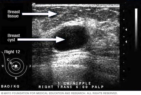
Ultrasound of breast cyst
This ultrasound shows a breast cyst.
Ultrasound of liver tumor
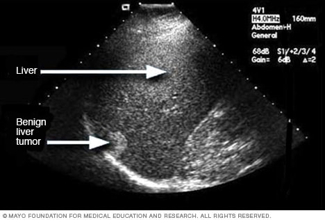
Ultrasound of liver tumor
An ultrasound uses sound waves to create an image. This ultrasound shows a noncancerous liver tumor.
Ultrasound of gallstones

Ultrasound of gallstones
This ultrasound shows gallstones in the gallbladder.
Ultrasound of needle-guided procedure
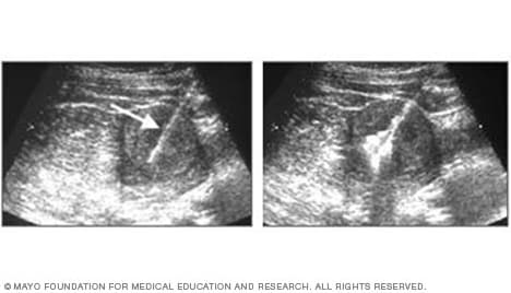
Ultrasound of needle-guided procedure
These images show how ultrasound can help guide a needle into a tumor (left), where material is injected (right) to destroy tumor cells.
Transvaginal ultrasound
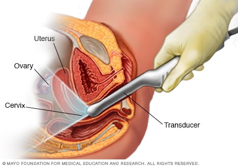
Transvaginal ultrasound
During a transvaginal ultrasound, a healthcare professional or technician uses a wandlike device called a transducer. The transducer is inserted into your vagina while you lie on your back on an exam table. The transducer emits sound waves that generate images of your pelvic organs.
Before your ultrasound begins, you may be asked to do the following:
- Remove any jewelry from the area being examined.
- Remove or reposition some or all of your clothing.
- Change into a gown.
You'll be asked to lie on an exam table.
During the procedure
Gel is applied to your skin over the area being examined. It helps prevent air pockets, which can block the sound waves that create the images. This safe, water-based gel is easy to remove from skin and, if needed, clothing.
A trained technician, called a sonographer, uses a small, hand-held device called a transducer. The technician presses the transducer against the area being studied and moves it as needed to capture the images. The transducer sends sound waves into your body and collects the ones that bounce back. The images appear on a computer.
Sometimes, ultrasounds are done inside the body. In this case, the transducer is attached to a probe that's inserted into a natural opening in the body. Examples include:
- Transesophageal echocardiogram. A transducer, inserted into the esophagus, obtains heart images. It's usually done while under sedation.
- Transrectal ultrasound. This test creates images of the prostate by placing a special transducer into the rectum.
- Transvaginal ultrasound. A special transducer is inserted into the vagina to look at the uterus and ovaries.
Ultrasound is usually painless. However, you may experience mild discomfort as the sonographer guides the transducer over your body. It may not be comfortable if you're required to have a full bladder or the transducer is inserted it into your body.
A typical ultrasound exam takes from 30 minutes to an hour.
Results
When your exam is complete, a doctor trained to interpret imaging studies, called a radiologist, analyzes the images. The radiologist sends a report to your healthcare professional who will share the results with you.
You should be able to return to usual activities right after an ultrasound.
Clinical trials
Explore Mayo Clinic studies of tests and procedures to help prevent, detect, treat or manage conditions.