Overview
Chest X-ray
Chest X-ray
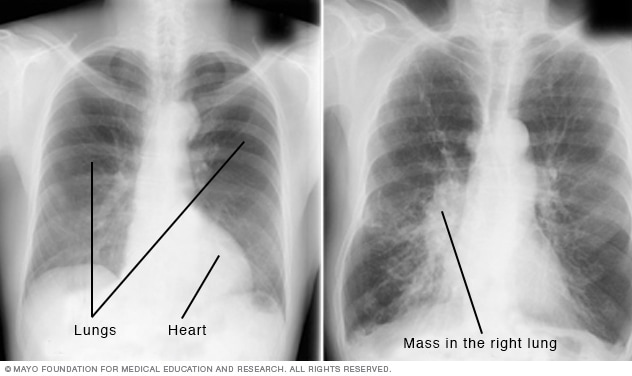
A chest X-ray helps detect concerns with your heart and lungs. The chest X-ray on the left does not show any issues. The image on the right shows a mass in the right lung.
Chest X-rays produce images of your heart, lungs, blood vessels, airways, and the bones of your chest and spine. Chest X-rays also can reveal fluid in or around your lungs or air surrounding a lung.
If you go to your healthcare professional or the emergency room with chest pain, a chest injury or shortness of breath, you will typically get a chest X-ray. The image helps your health professional find out if you have heart issues, a collapsed lung, pneumonia, broken ribs, emphysema, cancer or any of several other conditions.
Some people have a series of chest X-rays done over time to track whether a health condition is getting better or worse.
Why it's done
A chest X-ray is used to examine the structures inside the chest, including the lungs, heart, airways, blood vessels, and the bones of the chest and spine.
A chest X-ray may be done to:
- Evaluate symptoms such as shortness of breath, a persistent cough, chest pain or fever.
- Help diagnose or monitor lung conditions, including pneumonia, emphysema, lung cancer, or air or fluid around the lungs.
- Diagnose fractures of the collar bones, breastbone or ribs.
- Assess the heart and major blood vessels, including changes in heart size that may be linked to certain heart conditions.
- Check the position of medical devices placed in the chest, such as pacemakers or tubes.
Because chest X-rays are fast and widely available, they are particularly useful in emergency and urgent care settings.
More Information
Risks
You may be concerned about radiation exposure from chest X-rays, especially if you have them regularly. But the amount of radiation from a chest X-ray is low — even lower than what you're exposed to through natural sources of radiation in the environment.
Even though the benefits of an X-ray outweigh the risk, you may be given a protective apron if you need multiple images. Tell your healthcare professional if you're pregnant or might be pregnant. The procedure can be performed in a way that protects your abdomen from radiation.
How you prepare
Before the chest X-ray, you generally undress from the waist up and wear an exam gown. You'll need to remove jewelry from the waist up, too, since both clothing and jewelry can obscure the X-ray images.
What you can expect
During the procedure, your body is positioned between a machine that produces the X-rays and a plate that creates the image digitally or with X-ray film. You may be asked to move into different positions in order to get views from both the front and the side of your chest.
For the front view, you stand against the plate, hold your arms up or to the sides, and roll your shoulders forward. The X-ray technician may ask you to take a deep breath and hold it for several seconds. Holding your breath after inhaling helps your heart and lungs show up more clearly on the image.
For the side views, you turn and place one shoulder on the plate and raise your hands over your head. Again, you may be asked to take a deep breath and hold it.
Having X-rays taken is generally painless. You don't feel any sensation as the radiation passes through your body. If you have trouble standing, you may be able to have the exam while seated or lying down.
Results
A chest X-ray produces a black-and-white image that shows the organs in your chest. Structures that block radiation appear white, and structures that let radiation through appear black.
Your bones appear white because they are very dense. Your heart also appears as a lighter area. Your lungs are filled with air and block very little radiation, so they appear as darker areas on the images.
The images are typically examined by a healthcare professional trained to interpret X-rays and other imaging exams, called a radiologist. The radiologist looks for clues that may suggest if you have heart failure, fluid around your heart, cancer, pneumonia or another condition.
Someone on your healthcare team will talk with you about the results and what treatments or other tests or procedures may be necessary.
Sept. 30, 2026