April 26, 2024
Artificial intelligence (AI) is a cornerstone of Mayo Clinic's efforts to improve the diagnosis and treatment of glioma. One effort involves creating predictive regional maps of tumor characteristics, to guide individualized treatment.
Currently, clinical decision-making relies on MRI interpretation and tumor sampling, which fails to capture the full heterogeneity of glioma. That heterogeneity is a major challenge, as it contributes to treatment resistance and tumor recurrence.
Mayo Clinic researchers are obtaining multiple image-localized biopsy samples and pairing them with multiparametric MRI. "By connecting different regions of tumor biology to different imaging features, this project ultimately aims to predict the locations of a tumor's biological features from imaging alone," says Kristin R. Swanson, Ph.D., a mathematical oncologist at Mayo Clinic in Arizona.
The research is funded by the National Cancer Institute and built on Mayo Clinic's large and growing resource of image-localized biopsies, as described in a 2023 article in PLOS One. The work is a collaborative effort between the Mathematical Neuro-Oncology Laboratory, led by Dr. Swanson; and the Neurosimulation and Innovations Laboratory, led by Bernard R. Bendok, M.D., chair of Neurosurgery at Mayo Clinic's campus in Arizona.
Smarter brain imaging is needed to overcome the limited opportunities for glioma tissue collection. "We want to make the imaging more intelligent by feeding it biopsy information," Dr. Bendok says. "The AI system may recognize and enhance patterns that we don't even recognize now."
Answering key questions
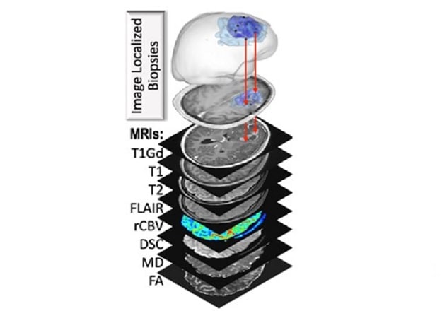 Multiparametric MRI techniques and contrasts
Mayo Clinic researchers pair image-localized biopsy samples with eight MRI sequences: T1Gd; T1-weighted; T2-weighted; fluid-attenuated inversion recovery (FLAIR); relative cerebral blood volume (rCBV); dynamic susceptibility contrast perfusion T2-W signal loss (DSC); diffusion-weighted imaging/diffusion tensor imaging mean diffusivity (MD); and diffusion-weighted imaging/diffusion tensor imaging fractional anisotropy (FA).
Patients who agree to participate in the research have multiple imaging studies before routine surgical intervention for a brain lesion. The imaging studies include dynamic susceptibility contrast perfusion MRI and diffusion tensor imaging, in addition to standard T1, T1Gd, T2 and T2 FLAIR scans.
During surgery, neuronavigation tracks sample anatomical locations. After surgery, genetic aberrations in the tissue samples are quantified with whole-exome and RNA sequencing, as well as other tissue analysis techniques. The resulting data are then used to generate, test and validate predictive regional maps of the spatial distribution of tumor cell density and treatment-related key genetic and other biological markers.
"We expect a large dataset of image-localized biopsies will help us answer some key questions surrounding the heterogeneity of gliomas," Dr. Swanson says.
The research already is shedding light on the biological complexity of infiltrative tumor in high- grade glioma. As described in Nature Communications, the researchers demonstrated that nonenhancing tumor regions harbor the highest proportion of private mutations, regardless of IDH status.
"That implies that mutational burden in high-grade glioma is subject to sample location," Dr. Swanson says. "We also propose that regional genomic instability and tumor infiltration occur early in the development of glioma."
Identifying biologically aggressive tumor regions can better inform future targeted treatment. "New therapies are urgently needed," Dr. Bendok says. "Diffusely invasive gliomas are particularly challenging, as there are always tumor cells left behind in the brain following any surgery."
Mayo Clinic's embrace of AI rests upon a commitment to patient care. "That desire requires us to take advantage of the digital revolution," Dr. Bendok says. "We value our strong traditions at Mayo Clinic. But every tradition was once a revolution."
For more information
Urcuyo JC, et al. Image-localized biopsy mapping of brain tumor heterogeneity: A single-center study protocol. PLOS One. 2023;18:e0287767.
Mathematical Neuro-Oncology Laboratory: Kristin R. Swanson. Mayo Clinic.
Neurosurgery Simulation and Innovations Laboratory: Bernard R. Bendok. Mayo Clinic.
Hu LS, et al. Integrated molecular and multiparametric MRI mapping of high-grade glioma identifies regional biologic signatures. Nature Communications. 2023;14:6066.
Refer a patient to Mayo Clinic.