Overview
Brain tumor MRI
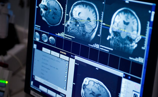
Brain tumor MRI
Brain tumor imaging
Magnetic resonance imaging (MRI) is a medical imaging technique that uses a magnetic field and computer-generated radio waves to create detailed images of the organs and tissues in your body.
Most MRI machines are large, tube-shaped magnets. When you lie inside an MRI machine, the magnetic field inside works with radio waves and hydrogen atoms in your body to create cross-sectional images — like slices in a loaf of bread.
The MRI machine also can produce 3D images that can be viewed from different angles.
Why it's done
MRI is a noninvasive way for a medical professional to examine your organs, tissues and skeletal system. It produces high-resolution images of the inside of the body that help diagnose a variety of conditions.
MRI of the brain and spinal cord
MRI is the most frequently used imaging test of the brain and spinal cord. It's often performed to help diagnose:
- Aneurysms of cerebral vessels.
- Conditions of the eye and inner ear.
- Multiple sclerosis.
- Spinal cord conditions.
- Stroke.
- Tumors.
- Brain injury from trauma.
A special type of MRI is the functional MRI of the brain, also known as fMRI. It produces images of blood flow to certain areas of the brain. Functional MRI can be used to examine the brain's anatomy and show which parts of the brain are handling critical functions, language and movements. This information can help guide decisions when considering someone for brain surgery.
Functional MRI also can check for damage from a head injury or from conditions such as Alzheimer's disease.
MRI of the heart and blood vessels
MRI that focuses on the heart or blood vessels can check:
- Size and function of the heart's chambers.
- Thickness and movement of the walls of the heart.
- Extent of damage caused by heart attacks or heart disease.
- Structural problems in the aorta, such as aneurysms or dissections.
- Inflammation or blockages in the blood vessels.
MRI of other internal organs
MRI can check for tumors or other irregularities in many organs in the body, including the following:
- Liver and bile ducts.
- Kidneys.
- Spleen.
- Pancreas.
- Uterus.
- Ovaries.
- Prostate.
MRI of bones and joints
MRI can help look for:
- Joint issues caused by traumatic or repetitive injuries, such as torn cartilage or ligaments.
- Disk problems in the spine.
- Bone infections.
- Tumors of the bones and soft tissues.
MRI of the breasts
MRI can be used with mammography to detect breast cancer, particularly in people who have dense breast tissue or who might be at high risk of the disease.
More Information
Risks
Because MRI uses powerful magnets, the presence of metal in your body can be a safety hazard if attracted to the magnet. Even if not attracted to the magnet, metal objects can distort the MRI images. Before having an MRI exam, you'll likely complete a questionnaire that includes whether you have metal or electronic devices in your body.
Unless the device you have is certified as MRI safe, you might not be able to have an MRI. Devices include:
- Metallic joint prostheses.
- Artificial heart valves.
- An implantable heart defibrillator.
- Implanted drug infusion pumps.
- Implanted nerve stimulators.
- A pacemaker.
- Metal clips.
- Metal pins, screws, plates, stents or surgical staples.
- Cochlear implants.
- A bullet, shrapnel or any other type of metal fragment.
- Intrauterine device.
If you have tattoos or permanent makeup, ask whether it might affect your MRI. Some of the darker inks contain metal.
Before you schedule an MRI, tell your doctor if you think you're pregnant. The effects of magnetic fields on an unborn baby aren't well understood. An alternative exam may be recommended, or the MRI may be postponed. Also tell your doctor if you're breastfeeding, especially if you're to receive contrast material during the procedure.
It's also important to discuss kidney or liver problems with your doctor and the technologist, because problems with these organs might limit the use of injected contrast agents during your MRI scan.
How you prepare
Before an MRI exam, eat as you would normally and continue to take your usual medicines, unless you're told otherwise. You will typically be asked to change into a gown and to remove things that might affect the magnetic imaging, such as:
- Jewelry.
- Hairpins.
- Eyeglasses.
- Watches.
- Wigs.
- Dentures.
- Hearing aids.
- Underwire bras.
- Cosmetics that contain metal particles.
What you can expect
During the test
The MRI machine looks like a long narrow tube that is open on both ends. You lie down on a movable table that slides into the opening of the tube. A technologist monitors you from another room. You can talk with the technologist by microphone.
If you have a fear of enclosed spaces, called claustrophobia, you might receive a drug to help you feel sleepy and less anxious. Most people get through the exam without difficulty.
The MRI machine creates a strong magnetic field around you, and radio waves are directed at your body. The procedure is painless. You don't feel the magnetic field or radio waves, and there are no moving parts around you.
During the MRI scan, the internal part of the magnet produces repetitive tapping, thumping and other noises. Wearing earplugs or having music playing can help block the noise.
In some cases, a contrast material, typically gadolinium, will be injected through an intravenous (IV) line into a vein in a hand or arm. The contrast material helps make certain details clearer. Gadolinium rarely causes allergic reactions.
An MRI exam can last anywhere from 15 minutes to more than an hour. You must hold still because movement can blur the images.
During a functional MRI exam, you might be asked to perform a few small tasks — such as tapping your thumb against your fingers, rubbing a block of sandpaper or answering simple questions. This helps pinpoint the portions of your brain that control these actions.
After the test
If you haven't been sedated, you can resume your usual activities immediately after the scan.
Results
A doctor specially trained to interpret MRI scans, called a radiologist, will look over the images from your scan and report the findings to your doctor. Your doctor will discuss important findings and next steps with you.
An MRI is a very useful tool for helping your doctors see images of the inside of your body, including tissue that can't be seen on a conventional x-ray.
Before your exam, it's very important to fill out the safety screening form carefully. MRI is safe and painless. But metal in the scanner can cause serious safety problems or reduce the quality of the images.
Your health care team needs to know about any metal in your body, even a small shard of metal from an accident. Fillings, bridges, and other dental work typically do not pose a problem. But other metal that has been put into your body might prevent you from having an MRI. That includes some pacemakers, clips for treating aneurysms, and other devices with metal in them.
A nurse may review your health history before your exam. You may be given medications or contrast dye or have blood drawn. Be sure to tell the nurse if you're pregnant, have an allergy to contrast dye, or have kidney or liver problems. You may not wear clothing with snaps or zippers in the scanner. You will be asked to wear a gown. Do not wear any jewelry or bring anything metal into the scanner, including a hearing aid.
An MRI machine uses a powerful magnet to make images of your body. Unlike a CT scan, it does not use x-rays or other radiation. You will be given earplugs. The scanner makes a loud noise when it's operating.
A device called a coil may be put on or around the area to be scanned to help capture the images. You will also be given a squeeze ball to hold. You can use this to signal the technologist any time you need something. The MRI is controlled from a nearby room. You will be closely observed throughout the procedure.
A series of scans are taken with a brief pause between each. You may hear different noises as different scans are taken. It's normal for the noise to be very loud. You need to remain still when the scan is being taken.
People are typically in the scanner from 30 to 50 minutes, depending on the images to be taken. A complex examination can take longer. If you are concerned about being in the scanner for this length of time, talk to your physician and the technologist. They can help you with some tips for staying comfortable.
If you need to be removed from the scanner, this can be done very quickly. The ends of the scanner are always open.
After your exam, the images will be reviewed by your radiologist. He or she will send a report to the health care provider who ordered the test. Ask your health care provider any questions you have about your MRI.
Seeing inside the heart with MRI
Watch how cardiac MRI uses still or moving pictures to show blood flow through the heart.
Vivien Williams: One out of four, that's how many people will die of a heart related problem. Doctors at Mayo Clinic are trying to improve those statistics. They're using MRIs to look inside the heart to find disease and tailor treatment to keep people healthier longer.
MRI technician: You can breathe. Breathe normal.
Vivien Williams: Magnetic Resonance Imaging, or MRI, allows doctors to look inside the heart as it beats.
Brian Shapiro, M.D., Mayo Clinic cardiologist: You can see here, this is the left ventricle, which is the main pumping chamber that pushes blood out of the body.
Vivien Williams: Dr. Brian Shapiro uses MRI to look for abnormalities in the heart.
Brian Shapiro, M.D.: What the MRI does is it looks at the tissue characteristics of the heart. So, swelling of the heart is a very common thing in heart attacks, and infections, and things like that.
Vivien Williams: The moving or still images show exactly where damage happens.
Brian Shapiro, M.D.: You would see it as a very bright, bright spot in the heart.
Vivien Williams: In addition to damage from heart attack or infection, MRI can also show Dr. Shapiro how well the heart pumps, where irregular heart beats originate, the location of blood clots, artery blockages, scar tissue, or even tumors. Because MRI allows doctors to see more detail of the heart, they can make more accurate diagnoses, and therefore tailor treatment for patients.
Brian Shapiro, M.D.: As you can actually show where the heart attack is, and the extent of the heart attack.
Vivien Williams: Images that tell Dr. Shapiro if a patient will recover, if there's permanent damage, and what treatments might be best. Information from inside the heart that can help Dr. Shapiro and his colleagues better help their patients. Dr. Shapiro says while MRI can show lots of information about the heart, it does not replace other tests such as stress tests or echo cardiograms. It's another option for looking inside the heart. For Medical Edge, I'm Vivien Williams.
Clinical trials
Explore Mayo Clinic studies of tests and procedures to help prevent, detect, treat or manage conditions.
Sept. 09, 2023