Overview
Epilepsy surgery is a procedure that removes an area of the brain where seizures occur.
Epilepsy surgery is most effective when seizures always occur in a single location in the brain. Epilepsy surgery is not the first line of treatment. But it might be an option when at least two anti-seizure medicines have failed to control seizures.
You might need several tests before surgery to find out whether epilepsy surgery is an option and what type of surgery to do.
Products & Services
Why it's done
Location of temporal lobe

Location of temporal lobe
The temporal lobe is located along each side of the brain.
Brain hemispheres
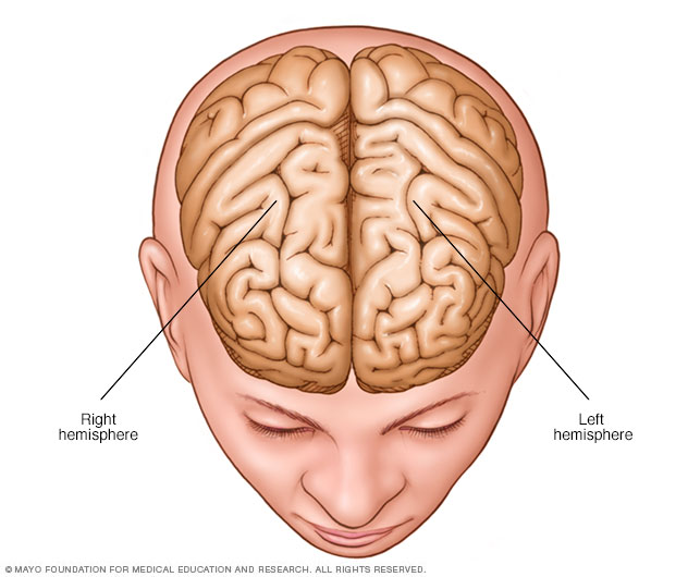
Brain hemispheres
The brain is divided into two halves, called hemispheres.
Epilepsy surgery may be an option when medicines don't control seizures. This is a condition known as medically refractory epilepsy or drug-resistant epilepsy. Epilepsy surgery is done to stop seizures or limit their severity. Surgery also is done to lessen seizure-related deaths, decrease the use of anti-seizure medications and decrease the possible side effects of the medicines.
Poorly controlled epilepsy can result in a number of complications and health risks, including:
- Physical injuries during a seizure
- Drowning, if the seizure occurs during a bath or swimming
- Depression and anxiety
- Developmental delays in children
- Sudden death, which is a rare complication of epilepsy
- Worsening memory or other thinking skills
Types of epilepsy surgery
Epileptic seizures result from irregular activity of brain cells called neurons. The type of surgery needed depends on the location of the neurons that start the seizure and the age of the person having the surgery. Types of surgery include:
- Resective surgery is the most common epilepsy surgery. It involves the removal of a small portion of the brain. The surgeon cuts out brain tissue from the area of the brain where seizures occur. This is usually the site of a tumor, brain injury or malformation. Resective surgery is usually performed on one of the temporal lobes. This is an area of the brain that controls visual memory, language comprehension and emotions.
- Laser interstitial thermal therapy (LITT) is less invasive than resective surgery. It uses a laser to pinpoint and destroy a small portion of brain tissue. Magnetic resonance imaging (MRI) is used to guide the laser.
- Deep brain stimulation is the use of a device that is placed permanently deep inside the brain. The device releases regularly timed electrical signals that disrupt seizure-inducing activity. This procedure is guided by MRI. The generator that sends the electrical pulse is implanted in the chest.
- Corpus callosotomy is surgery to completely or partially remove the part of the brain that connects nerves on the right and left sides of the brain. This part of the brain is called the corpus callosum. This surgery is usually used with children who experience irregular brain activity that spreads from one side of the brain to the other.
- Hemispherectomy is a procedure to remove one side of the brain called the cerebral cortex. This surgery is generally done only in children who experience seizures that originate from multiple sites in one hemisphere, usually the result of a condition present at birth or in early infancy.
- Functional hemispherectomy is a procedure primarily used in children that removes the connecting nerves without removing actual pieces of the brain.
Risks
Different areas of the brain control different functions. The risks vary depending on the surgical site and the type of surgery. The health care team explains the specific risks of the procedure, as well as the ways they can reduce the risk of complications. Risks may include:
- Memory and language problems that can affect the ability to understand and use language
- Visual impairment where the fields of vision of the eyes overlap
- Depression or other mood changes that can affect social relationships
- Headache
- Stroke
How you prepare
If someone is a possible candidate for epilepsy surgery, that person works with a health care team at a specialized epilepsy center. The health care team does several tests to:
- Determine the eligibility for surgery
- Identify the appropriate surgical site
- Understand in detail how that area of the brain functions
Some of these tests are done as outpatient procedures, while others require a hospital stay.
Evaluations to find the problem area
Pinpointing seizure location
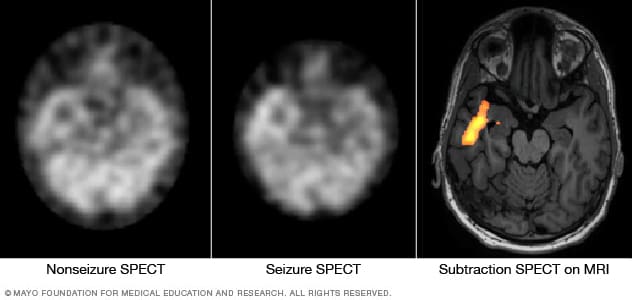
Pinpointing seizure location
These SPECT images show the blood flow in the brain of a person when there's no seizure activity (left) and during a seizure (middle). The subtraction SPECT coregistered to MRI (right) helps pinpoint the area of seizure activity by overlapping the SPECT results with brain MRI results.
These procedures are standard tests used to identify the source of irregular brain activity.
- Baseline electroencephalogram (EEG). In this test, electrodes are placed on the scalp to measure electrical activity produced by the brain when a seizure is not present. This test can suggest general areas of the brain that may be affected and can be done while asleep or awake.
- Video EEG. A continuous EEG with video monitoring records seizures as they occur. Because seizure medicines have to be reduced or temporarily stopped so that seizures will occur, this test is done in the hospital. Evaluating the changes in the EEG with body movements during a seizure helps pinpoint the area of the brain where the seizures are starting.
- Magnetic resonance imaging (MRI). This imaging test uses a magnetic field and radio waves to create detailed images that allow health care providers to identify damaged cells, tumors or other irregularities that can cause seizures.
The surgical team may order additional tests to localize the source of seizures and to characterize the nature of the unusual activity. These tests may include:
- Invasive EEG monitoring. If an EEG test does not show where seizures begin, monitoring may be done with surgically placed electrodes. The surgeon places either grids or strips of electrodes on the surface of the brain or places electrodes deeper inside the brain. EEG monitoring is done while the person is unconscious.
- Video EEG with invasive electrodes. Surgically placed electrodes may also be needed for a video EEG procedure. After the surgery, the video and EEG data are captured during a hospital stay while the person is awake but not taking anti-seizure medications.
- Positron emission tomography (PET). This specialized imaging device is used to measure brain function when the person is seizure-free. The images alone — or combined with MRI data — can help identify the source of the seizures.
- Single-photon emission computerized tomography (SPECT). This procedure measures blood flow in the brain during a seizure. Typically, blood flow is higher in the part of the brain where seizures occur. People are admitted to the hospital to undergo this test.
Evaluations to understand brain function
Depending on the surgical site, the health care team may recommend tests to determine the precise areas of the brain that control language, sensation, motor skills and other important functions. This information helps the surgeon preserve function to the greatest extent possible when removing or altering a site in the brain.
The tests may include the following:
- Functional MRI. This test identifies regions of brain activity when someone is doing a particular task, such as listening or reading. This helps the surgeon know the precise locations in the brain that control a particular function.
- Wada test. With this test, an injected medicine temporarily puts one side of the brain to sleep at a time. Then a test for language and memory function is done. This test can help determine which side of the brain is dominant for language usage. While functional MRI has often replaced this test, it may be used if the imaging study isn't possible.
- Brain mapping. Small electrodes are surgically placed on the surface of the brain. When the person is alert after the surgery, a number of tasks are performed that are matched with measurements of the brain's electrical activity.
- Magnetoencephalography (MEG). This test analyzes the magnetic fields produced by the electrical currents in the brain. It is used with data from other sources to locate seizure sites.
Neuropsychological tests
Additionally, testing is usually recommended to measure verbal and nonverbal learning skills and memory function. These tests may provide additional insight into the area of the brain affected by seizures, as well as a baseline for measuring function after surgery.
What you can expect
Before the procedure
To avoid infection, hair is clipped short or shaved over the section of the skull that will be removed during the operation. A small, flexible tube is placed within a vein to deliver IV fluids, anesthetics or other medicines during the surgery.
During the procedure
Heart rate, blood pressure and oxygen levels are monitored throughout the surgery. An EEG monitor may record brain waves during the operation to better localize the part of the brain where seizures start.
Epilepsy surgery is usually done using general anesthesia. The person is unconscious during the procedure. In rare circumstances, the surgeon may awaken the person during part of the operation to help the team determine which parts of the brain control language and movement. In such cases, medicine is used to control pain.
The surgeon creates a relatively small window in the skull, depending on the type of surgery. After surgery, the bone is replaced and fastened to the remaining skull for healing.
After the procedure
The person is placed in a special recovery area to be monitored carefully after awakening from anesthesia. The person is usually placed in the intensive care unit the first night after surgery. The total hospital stay for most epilepsy surgeries is usually about three or four days.
After awakening, the head will be swollen and painful. Most people need to take pain medicine for at least the first few days. An ice pack on the head also may help. Most postoperative swelling and pain resolve within several weeks.
Most people are not able to return to work or school for about 1 to 3 months. Rest and relaxation are needed for the first few weeks after epilepsy surgery and then physical activity can be increased.
It's unlikely that intensive rehabilitation will be needed as long as the surgery was completed without complications such as a stroke or loss of speech.
Results
The outcomes of epilepsy surgery vary depending on the type of surgery performed. The expected outcome is seizure control with medication.
The most common and best-understood procedure — resection of tissue in the temporal lobe — results in seizure-free outcomes for about two-thirds of people. Studies suggest that if a person takes seizure medicine and does not have a seizure in the first year after temporal lobe surgery, the likelihood of being seizure-free at two years is 87% to 90%. If there are no seizures in two years, the likelihood of being seizure-free is 95% at five years and 82% at 10 years.
If there are no seizures for at least one year, the health care provider may consider decreasing the anti-seizure medicine and eventually discontinuing it entirely. Most people who do experience a seizure after going off medicine are able to obtain seizure control by resuming medicine.
Clinical trials
Explore Mayo Clinic studies of tests and procedures to help prevent, detect, treat or manage conditions.
Oct. 21, 2022