Overview
Carotid artery
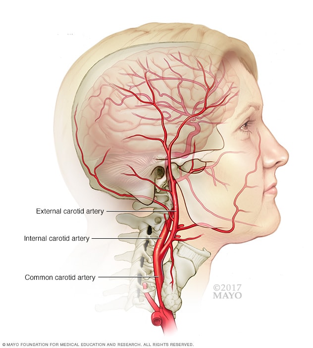
Carotid artery
The carotid arteries are a pair of blood vessels. There's one on each side of the neck. The carotid arteries deliver blood to the brain and head.
Carotid (kuh-ROT-id) ultrasound is a safe, noninvasive, painless procedure that uses sound waves to examine the blood flow through the carotid arteries. It also evaluates the thickness of the carotid artery wall and checks for clots.
One carotid artery is located on each side of the neck. These arteries deliver blood from the heart to the brain.
A carotid ultrasound tests for blocked or narrowed carotid arteries, which can increase the risk of stroke. The results of the test can help your health care provider determine a treatment to lower your stroke risk.
Products & Services
Why it's done
Ischemic stroke
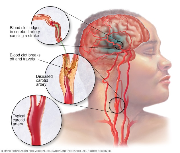
Ischemic stroke
An ischemic stroke occurs when a blood clot, known as a thrombus, blocks or plugs an artery leading to the brain. A blood clot often forms in arteries damaged by a buildup of plaques, known as atherosclerosis. It can occur in the carotid artery of the neck as well as other arteries.
A carotid ultrasound is done to look for for narrowed carotid arteries, which increase the risk of stroke.
Carotid arteries are usually narrowed by a buildup of plaque — made up of fat, cholesterol, calcium and other substances that circulate in the bloodstream. Early diagnosis and treatment of a narrowed carotid artery can decrease stroke risk.
Your health care provider orders a carotid ultrasound if you have a transient ischemic attack (TIA), also called a mini-stroke, or other types of strokes. Your provider also may recommend a carotid ultrasound if you have a medical condition that increases the risk of a stroke, including:
- High blood pressure
- Diabetes
- High cholesterol
- Family history of stroke or heart disease
- Recent transient ischemic attack (TIA) or stroke
- Unusual sound in the carotid arteries (bruit) detected using a stethoscope
- Coronary artery disease
- Hardening of the arteries
Other uses of carotid ultrasound
Your doctor may order a carotid ultrasound to:
- Evaluate blood flow through the artery after surgery to remove plaques. The procedure to remove plaques is called carotid endarterectomy.
- Evaluate the placement and effectiveness of a stent, a mesh tube used to improve blood flow through an artery.
- Locate a collection of clotted blood, also called a hematoma, that may prevent blood flow.
- Detect other carotid artery problems that may disrupt blood flow.
- Predict coronary artery disease by measuring the thickness of the carotid artery and evaluating the characteristics of a plaque.
- Monitor carotid artery blood flow during aortic heart valve surgery to assess the risk of a stroke.
- Construct a 3D model of the carotid artery to improve the accuracy of a diagnosis.
More Information
How you prepare
You can take the following steps to prepare for your appointment:
- Call the day before the exam to confirm the time and location of the exam.
- Wear a comfortable shirt with no collar or an open collar.
- Don't wear a necklace or dangling earrings.
Unless your health care provider or the radiology lab provides special instructions, you shouldn't need to make any other preparations.
What you can expect
How it works
A technician called a sonographer conducts the test with a small, hand-held device called a transducer. The transducer emits sound waves and records the echo as the waves bounce off tissues, organs and blood cells.
A computer translates the echoed sound waves into a live-action image on a monitor. The ultrasound technician may use a Doppler ultrasound, which shows blood flowing through the arteries. In a Doppler ultrasound, the rate of blood flow is translated into a graph.
There have been vast technological advances in carotid ultrasounds, improving the quality and resolution of the images.
A carotid ultrasound usually takes about 30 minutes.
During the procedure
You'll likely lie on your back during the ultrasound. The ultrasound technician may position your head to better access the side of your neck.
The ultrasound technician will apply a warm gel to your skin above the site of each carotid artery. The gel helps transmit the ultrasound waves back and forth. The technician then gently presses the transducer against the side of your neck.
You shouldn't feel any discomfort during the procedure. If you do, tell the ultrasound technician.
Results
A doctor who specializes in imaging tests, called a radiologist, will review your test results, then prepare a report for the health care provider who ordered the test. This may be your health care provider, a doctor trained in heart and blood vessel conditions, called a cardiologist, or a doctor trained in brain and nervous system conditions, called a neurologist.
The radiologist also may discuss the results of the test with you immediately after the procedure.
The health care provider who ordered the test will explain to you what the carotid ultrasound revealed and what that means for you.
If the test shows that you're at risk of a stroke, your health care provider may recommend the following therapies depending on the severity of the blockage in your arteries:
- Eat a healthy diet, including fruits, vegetables, and whole-grain breads and cereals, and limit saturated fat.
- Exercise regularly.
- Keep a healthy weight.
- Eat a heart-healthy diet such as the Mediterranean diet
- Don't smoke, and avoid secondhand smoke.
- Take medications to lower blood cholesterol and blood pressure.
- Take medications to prevent blood clots.
- Have a surgical procedure to remove carotid artery plaques. This procedure is called carotid endarterectomy.
- Have a surgical procedure to open and support your carotid arteries. This procedure is called carotid angioplasty and stenting.
If your health care provider ordered the carotid ultrasound as a follow-up to a surgical procedure, your provider can explain whether the treatment is working and whether you'll need additional treatment or follow-up exams.
Additional tests
If your results are unclear, you may have additional imaging tests, including:
- A computerized tomography angiogram (CTA) scan. A CTA scan uses a series of X-rays to produce detailed images of the blood vessels in the body. A dye may be injected into a vein to make the carotid arteries easier to see on the scan.
- Magnetic resonance imaging (MRI). An MRI uses a magnetic field and radio waves to produce detailed images of soft tissues in the body. A magnetic resonance angiography (MRA) scan also may be performed to get a better look at blood vessels.
- Contrast-enhanced ultrasound (CEUS). An intravenous contrast agent helps find early hardening of the arteries and evaluate future vascular disease.
- 3D ultrasound. This test improves the visualization of blood vessels and helps in evaluating plaque progression, but it may underestimate narrowing of the carotid artery.
Jan. 12, 2023