Overview
Spinal arteriovenous malformation (AVM)
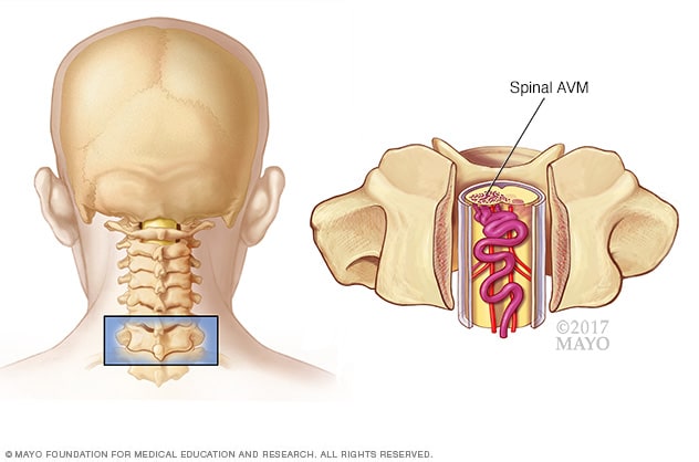
Spinal arteriovenous malformation (AVM)
A spinal arteriovenous malformation (AVM) is a tangle of blood vessels on, in or near the spinal cord.
A spinal arteriovenous malformation (AVM) is a tangle of blood vessels that forms on, in or near the spinal cord. This creates irregular connections between arteries and veins. Without treatment, this rare condition can cause lasting damage to the spinal cord.
Oxygen-rich blood enters the spinal cord through arteries. The arteries usually branch into smaller blood vessels called capillaries. The spinal cord gets oxygen from the blood in the capillaries. Then the blood passes into veins and moves away from the spinal cord to the heart and lungs.
But in a spinal AVM, the blood passes directly from the arteries to the veins. This change in blood flow means that the surrounding cells don't get the oxygen they need. This can cause cells in the spinal tissue to weaken or die.
The tangled arteries and veins in a spinal AVM also can burst and cause bleeding in the spinal cord. Sometimes, the AVM gets bigger as blood flow increases. The AVM can press on the spinal cord and cause weakness or other symptoms.
You might not know you have a spinal AVM unless you have symptoms. The condition can be treated with surgery to stop or possibly reverse some of the spinal damage.
Products & Services
Symptoms
Symptoms of a spinal arteriovenous malformation (AVM) can differ from person to person. Symptoms depend on where the AVM is found and how serious it is. Some people may not notice symptoms for many years, if at all. Others may experience symptoms that are life-threatening.
Symptoms often begin when people are in their 20s but can occur at earlier or later ages. Some people are diagnosed under the age of 16.
Symptoms may start suddenly or slowly and may include:
- Trouble walking or climbing stairs.
- Numbness, tingling or sudden pain in the legs.
- Weakness on one or both sides of the body.
As the condition gets worse, you may have more symptoms including:
- Sudden, serious back pain.
- Lack of feeling in the legs.
- Trouble urinating or having bowel movements.
- Headache.
- Stiff neck.
When to see a doctor
Make an appointment with your healthcare professional if you experience symptoms of a spinal AVM.
Causes
The cause of spinal arteriovenous malformations (AVMs) isn't known. Most spinal AVMs are present at birth, known as congenital. But others may happen later in life.
Risk factors
There are no known risk factors for spinal arteriovenous malformations (AVMs). The condition occurs equally in men and women.
Complications
Without treatment, a spinal arteriovenous malformation (AVM) can cause disability that gets worse over time. This is from damage to the spinal cord and surrounding tissues. This can cause:
- Trouble moving.
- Pain, tingling and numbness.
- Damage to the spine.
- Bulging blood vessel, known as an aneurysm.
- High blood pressure in the veins, known as venous hypertension. This can cause fluid to build up, called edema. It also can cause tissues to die due to lack of oxygen, known as spinal cord infarction.
- Hemorrhage, which can speed up spinal cord damage.