March 28, 2017
Zenker's diverticulum (ZD) results from posterior herniation of esophageal mucosa into Killian's triangle, an area of least resistance situated above the cricopharyngeus (CP) muscle and below the inferior pharyngeal constrictor muscle. ZD is actually a pseudodiverticulum since the Zenker's pouch contains only esophageal mucosa and submucosa. ZD can cause dysphagia and the pooling of food within the diverticulum, which can cause frequent regurgitation and can lead to aspiration pneumonia.
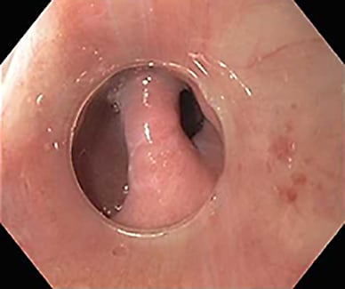
Zenker's diverticular pouch, post-cricopharyngeal myotomy
Zenker's diverticular pouch, post-cricopharyngeal myotomy
Zenker's diverticular pouch, post-cricopharyngeal myotomy with severing of the septum
Zenker's diverticular pouch
Zenker's diverticular pouch
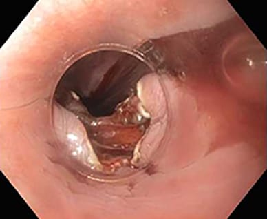
Zenker's diverticular pouch (left) and septum (center) relative to esophageal lumen (right)
Treatment for symptomatic ZD can be surgical or endoscopic. The surgical approach involves an external neck incision with CP myotomy (diverticulotomy), with or without pouch intervention (inversion, diverticulopexy or diverticulectomy). The endoscopic approach, using rigid or flexible endoscopes, involves only a diverticulotomy, in which the septum between the esophageal lumen and the diverticulum and the CP muscle are severed to create a single channel.
Each of these treatment approaches has variations in techniques and associated advantages and disadvantages. In an article published in Clinical Gastroenterology and Hepatology in 2014, the authors noted that although the open surgical approach offers effective symptom resolution in 90 to 95 percent of patients, its complication rate approximates 10 percent. Potential adverse events include fistula formation, abscess, hematoma, recurrent laryngeal nerve paralysis, difficulties in phonation and Horner's syndrome.
The endoscopic approach using rigid endoscopes also offers symptom relief in at least 90 percent of patients, with a 7 to 8 percent risk of adverse events, which include dental injury and perforation. However, the rigid endoscope approach cannot be performed in all patients, as in those with upper teeth protrusion, inadequate jaw opening or limited neck mobility that precludes adequate rigid endoscopic exposure of the ZD. The use of a rigid diverticuloscope also increases the risk of perforation when there is insufficient protection of a small diverticular pouch by the dorsal esophageal wall.
Benefits of the flexible endoscopic approach
Introduced nearly 20 years ago, flexible endoscopic therapy for ZD is becoming more widely accepted as newer data show sustained efficacy that is comparable to open surgical and rigid endoscopic therapies. In a systematic review and meta-analysis published in Gastrointestinal Endoscopy in 2016, the authors reported that the pooled success, adverse events and recurrence rates were 91 percent, 11.3 percent and 11 percent, respectively. Adverse events include post-procedural throat pain, bleeding and perforation.
In another article with similar study design published in Endoscopy International Open in 2016, the endoscopic approach appeared to result in a shorter length of procedure and hospital stay, earlier diet resumption and lower rates of adverse events, but in higher rates of symptom recurrence relative to the surgical approach.
Flexible endoscopy overcomes some of the limitations associated with the use of rigid instruments. The endoscope's flexibility and smaller diameter make this approach especially useful in patients with poor neck extension and/or limited jaw opening. In some instances, the procedure can be performed without the use of general anesthesia, and this approach may be preferable in patients with comorbid medical conditions that hinder surgical intervention.
Techniques of the flexible endoscopic approach
During flexible endoscopic myotomy, the septum (CP muscle) can be divided using a standard endoscopic retrograde cholangiopancreatography (ERCP) needle knife, as well as a HookKnife or HybridKnife, which are electrosurgical knives commonly used during endoscopic submucosal dissection. Additional devices to sever the septum have been reported, including monopolar and bipolar forceps, argon plasma coagulation and endoscopic scissors. Technical variations, including volumetric resection and submucosal tunneling for CP myotomy, also have been described.
A high level of technical expertise is required to perform this procedure. The main challenge is determining the depth of the CP myotomy needed to treat the condition adequately. An insufficient myotomy will result in ongoing symptoms, whereas too deep a resection will result in a perforation. As cutting techniques continue to evolve, comparative trials will be needed to establish which methods optimize outcomes in terms of efficacy and safety.
Mayo Clinic gastroenterologists have gained significant experience performing this procedure using a flexible endoscope. To date, the Mayo Clinic experience and recently published data suggest that one or two treatments using the flexible endoscopic approach can provide a high rate of symptom relief with a good safety profile, as well as a low rate of diverticular recurrence or persistence.
"The flexible endoscopic approach has demonstrable advantage over rigid endoscopy by way of control, ease and therapeutic options," says Timothy A. Woodward, M.D., a gastroenterologist specializing in endoscopic procedures at Mayo Clinic's campus in Florida. "Mayo has become one of the referral centers for the Southeastern United States in this regard," adds Dr. Woodward.
Louis M. Wong Kee Song, M.D., a therapeutic endoscopist who performs this procedure at Mayo Clinic's campus in Minnesota, notes that he and his colleagues typically perform 10 to 12 such procedures a year. "The flexible endoscopic treatment of Zenker's diverticulum is a niche practice given the specific skills needed for its performance and the uncommon occurrence of the condition."
Norio Fukami, M.D., a director of therapeutic endoscopy at Mayo Clinic's campus in Arizona, concurs that use of the flexible endoscopic approach is evolving as a newer option for the treatment of symptomatic Zenker's diverticulum. "We offer flexible endoscopic ZD myotomy with modified technique, aiming for higher efficacy and fewer adverse events. Staff at Mayo's three sites will continue collaborating to innovate in how we approach ZD myotomy, in a minimally invasive fashion with higher success rates."
For more information
Law R, et al. Zenker's diverticulum. Clinical Gastroenterology and Hepatology. 2014;12:1773.
Ishaq S, et al. Flexible endoscopic treatment for Zenker's diverticulum: A systematic review and meta-analysis. Gastrointestinal Endoscopy. 2016;83:1076.
Albers DV, et al. Endoscopic versus surgical approach in the treatment of Zenker's diverticulum: Systematic review and meta-analysis. Endoscopy International Open. 2016;4:E678.